PROCEDURE 43 Open Reduction and Internal Fixation of Sacral Fractures
Examination/Imaging
 Advanced Trauma Life Support guidelines are useful for identifying and managing injuries in a prioritized fashion.
 A detailed neurologic examination is important to identify associated neurologic injuries, but may not be definitive in the setting of the obtunded or multiply injured patient.
 Pelvic inlet and outlet views and a lateral radiograph of the sacrum may also add valuable information.
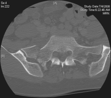
 Computed tomography (CT) scanning with or without three-dimensional reconstructions can be very helpful in understanding the injury pattern and planning treatment. The CT scan in Figure 1 demonstrates a displaced transforaminal fracture of the right sacral ala.
Surgical Anatomy
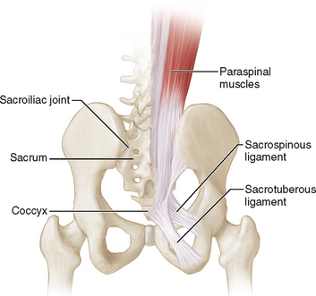
 The sacrum consists of five fused sacral vertebrae (unless there is a transitional segment) and the coccyx (Fig. 2)
• The anterior sacroiliac ligaments are relatively weak compared to the posterior ligamentous complex.
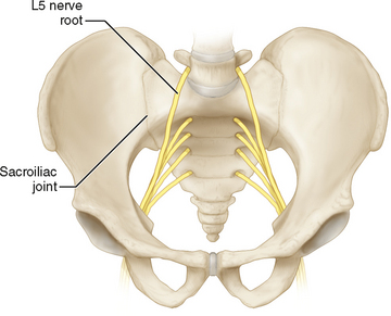
 Neural elements (Fig. 3)
• The neural elements travel in the spinal canal and branch off bilaterally through the sacral foramina (S1-4).
 The rich blood supply, the presacral plexus, and the proximity of the rectum limit access to the sacrum anteriorly in the midline.
 The paraspinal musculature attaches along the medial sacral crest and posterior iliac crest (see Fig. 2).
• Internal degloving injuries increase the risk of wound complications significantly and may dictate concomitant débridement or selection of an alternate approach.


