PROCEDURE 42 Open Reduction and Internal Fixation of Intra-articular Iliac Fracture-Subluxation (Crescent Fracture)
Indications
Examination/Imaging
 Initial resuscitation of the patient should be conducted as per Advance Trauma Life Support guidelines as these injuries are often associated with high-energy trauma with or without multiple injuries.
 The L5 nerve root and sciatic nerve are particularly at risk with fracture-dislocations. Figure 1 shows a fracture-dislocation of the right ilium with medial displacement and L5 nerve root injury.
 Inspection of the soft tissue envelope must be done to identify areas of concern that may influence choice of surgical approach, timing, and management.
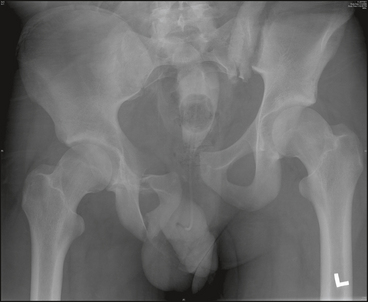
 Initial imaging includes an anteroposterior (AP) radiograph of the pelvis as well as inlet and outlet views. The AP pelvis radiograph in Figure 2 shows a transarticular (crescent) fracture of the left ilium.
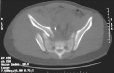
 Computed tomography (CT) scanning with or without three-dimensional reconstructions is often helpful in understanding fracture pattern and in preoperative planning. The CT scan in Figure 3 demonstrates comminution and extension into the sacroiliac (SI) joint.
Surgical Anatomy
 Intra-articular iliac fracture
• An intra-articular iliac fracture extends into the SI joint. It usually represents a lateral compression injury with rotational instability
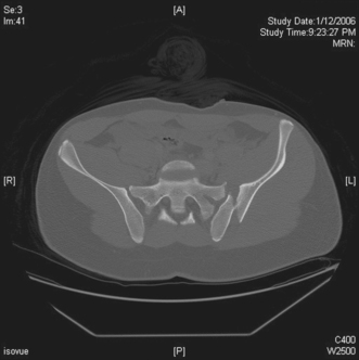
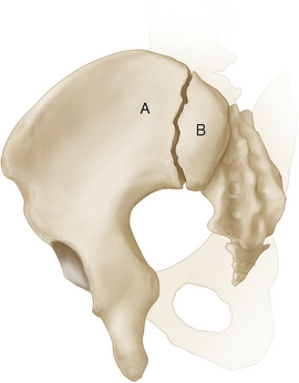
• The posterior superior iliac spine (PSIS) remains firmly attached to the sacrum via the strong posterior iliosacral ligaments. Figure 4 shows a transarticular fracture of the ilium with displacement of the anterior portion of the ilium and SI joint (A). The posterior fragment (B) generally remains well fixed and stable due to the strong posterior iliosacral ligaments.
 Structures at risk
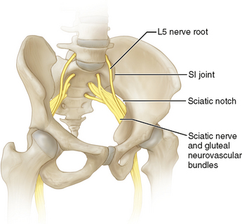
• Pelvic nerves (Fig. 5)
♦ The L5 nerve root travels over the ala of the sacrum anteriorly, approximately 2.5 cm medial to the SI joint.
