PROCEDURE 41 Anterior Pelvic Internal Fixation
Indications
 Pelvic injuries with symphyseal disruption or pubic body fractures (typically resulting in symphyseal diastasis of greater than 2.5 cm on an anteroposterior radiograph of the pelvis).
 These “open book” pelvic injuries may require simultaneous posterior fixation of the sacroiliac joint.
Examination/Imaging
 These injuries are usually the result of high-energy trauma, and patients should be evaluated for other simultaneous injuries and hemodynamic instability. Evaluation and resuscitation should follow Advanced Trauma Life Support principles.
 Specific other injuries should be suspected and managed appropriately.
• Pelvic fractures can result in significant urologic, vaginal, rectal, intra-abdominal, and neurovascular injuries.
 The condition of the soft tissue envelope needs to be assessed carefully for open injuries, including rectal and vaginal injuries, and severe crush injuries, as these will alter management.
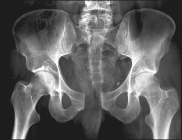
 Imaging should include an anteroposterior radiograph of the pelvis (Fig. 1) and inlet and outlet radiographs of the pelvis.
Surgical Anatomy
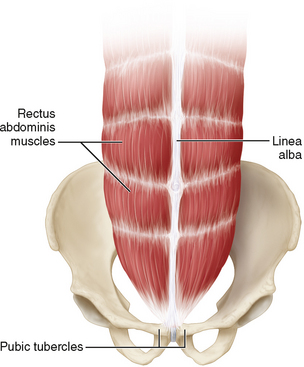
 Muscular anatomy (Fig. 2)
• The anterior rectus sheath is thick anteriorly but is of variable thickness posteriorly below the level of the anterior superior iliac spine, and care must be taken during dissection to stay extraperitoneal.
• The rectus abdominis muscle lies within the rectus sheath. The linea alba lies in the midline of the rectus abdominis. It provides an avascular plane for dissection and provides good tissue for closure.
• The insertion of the rectus abdominis is often avulsed on the side of the injury, and this defect should be incorporated into the exposure to minimize further stripping of the muscle insertion.
• The dissection is continued laterally past the pubic tubercles by 1 cm on each side, preserving as much of the anterior insertion of the rectus abdominis as possible.
• The patient should be placed truly level on the table with no twisting or rotation as this helps ensure accurate reduction.
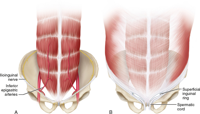
 Neurovascular anatomy (Fig. 3A)
• The inferior epigastric arteries also cross the surgical field and require ligation or cauterization.
 Fixation of the mid-position of the superior pubic ramus is possible through this approach, but fractures of the lateral portion of the superior pubic ramus require formal extension to an ilioinguinal approach or percutaneous screw fixation.
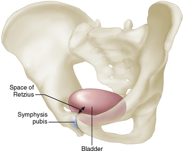
 The bladder lies posterior to the symphysis pubis with a potential space (of Retzius) between the two (Fig. 4).
• This space can be opened by blunt dissection with a finger; however, care needs to be exercised if there has been previous surgery in this region and/or a delay in fixation as adhesions may form between the pubic bone and the bladder.
