PROCEDURE 40 Pelvic External Fixation
Indications
 Temporary stabilization of pelvis fractures in hemodynamically unstable patients as part of resuscitation


Examination/Imaging
• Will a sheet/binder work as well as external fixation in the unstable patient and be quicker and easier to apply?
• Pelvic sheeting/binder in hemodynamically unstable patients with external rotation and vertically unstable fractures
• Internally rotating and strapping together the lower extremities in hemodynamically unstable patients with external rotation and vertically unstable fractures
• Pelvis C clamp in hemodynamically unstable patients with external rotation and vertically unstable fractures
• Open reduction and internal fixation or percutaneous fixation of pubic rami fractures and/or symphyseal diastasis as definitive treatment of the anterior pelvis injury
 Gentle physical examination is performed to determine internal/external rotational instability and vertical instability.
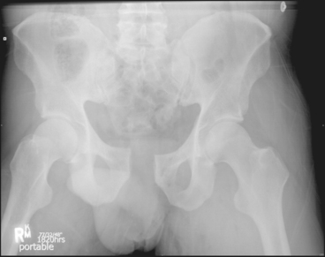
 An anteroposterior (AP) pelvis radiograph is usually sufficient in hemodynamically unstable patients.
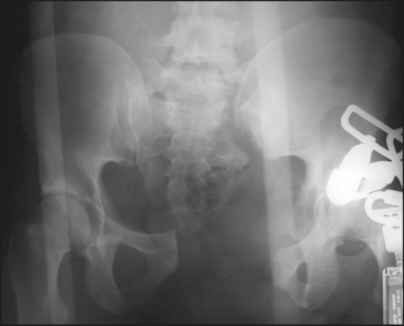
• Figure 1 shows a vertically unstable fracture (type C2) in a hemodynamically unstable polytrauma patient (Case 1).

Surgical Anatomy
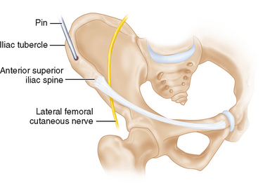
 Pin placement in the iliac crest insertion between the anterior superior iliac spine (ASIS) and iliac tubercle (Fig. 4)
• The curvature of the crest and obliquity of the ilium must be taken into consideration so that the pins converge in the dense bone proximal to the acetabulum.
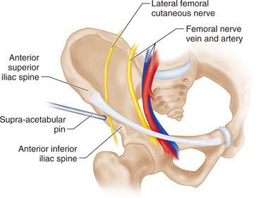
 Supra-acetabular (anterior) pin placement between the ASIS and anterior inferior iliac spine (AIIS) (Fig. 5)