This 50-year-old woman presents with patchy loss of pigment over her feet and hands (Fig. 24.1 and Fig. 24.2).
1 What is the most likely cause of hypopigmentation of the feet and hands?
2 Are there any underlying diseases associated with hypopigmentation?
3 What other conditions may lead to loss of skin pigment and why is it a cause for concern in some countries?
4 What precautions should this lady be aware of?
5 Can it be treated?
 |
| Fig. 24.1 |
Vitiligo
2 The cause is thought to be autoimmune disease with antimelanocyte antibodies. Organ-specific autoimmune diseases such as Hashimoto’s thyroiditis, Addison’s hypoadrenalism, pernicious anaemia and diabetes are associated with vitiligo. Melanocytes are absent from the white lesions on histology.
3 Albinism and phenylketonuria cause generalized hypopigmentation whereas leprosy, tinea versicolor and lichen sclerosis result in patchy loss of skin pigment. Vitiligo can be mistaken for the depigmented patches of Hansen’s disease (leprosy) and sufferers often become social outcasts.
4 The vitiliginous areas become easily sunburned and high factor sunscreen is required in the summer months.
5 Treatment can be unsatisfactory. Re-pigmentation can be achieved with exposure to long-wave ultraviolet light (UVA) together with psoralen (PUVA). Recent evidence shows that narrow band UVA (NB-UVA) is more effective than PUVA. The application of potent steroids can also induce re-pigmentation. Sunscreens reduce tanning of the pigmented skin and therefore reduce the contrast in skin tones. Cosmetic cover-up and self-tanning preparations can also be used.
A systematic review of 19 RCTs revealed that potent topical steroids resulted in better repigmentation than placebo and they were also better than oral psoralens plus sunlight in another study. Two studies suggested that topical calcipotriol enhanced repigmentation rates from PUVAsol and PUVA when compared with placebo.
Key points
• The cause is thought to be autoimmune disease with antimelanocyte antibodies.
• Vitiliginous areas become easily sunburned.
• Re-pigmentation can be achieved with exposure to long-wave ultraviolet light (UVA) together with psoralen (PUVA).
Further reading
Lim, HW; Hexsel, CL, Vitiligo: to treat or not to treat, Archives of Dermatology 143 (2007) 643–646.
Whitton, ME; Ashcroft, DM; Barrett, CW; Gonzalez, U, Interventions for vitiligo, Cochrane Database of Systematic Reviews (Issue 1) (2006).
Yones, S; Palmer, R; Garibaldinos, T; Hawk, J, Randomized double-blind trial of treatment of vitiligo: efficacy of psoralen-UV-A therapy vs narrowband-UV-B therapy, Archives of Dermatology 143 (2007) 578–584.
Case 25
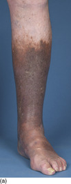
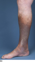
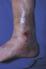
This 65-year-old man has discolouration of the skin over his lower leg and medial ankle (Fig. 25.1). He has been troubled by a recurrent ulcer in this area (Fig. 25.2).
1 What is the underlying pathology?
2 Explain the cause of the discolouration seen.
3 What management is appropriate?
 |
| Fig. 25.2 |
Venous ulceration
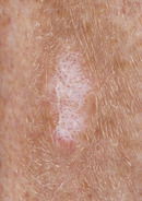
2 The extensive discolouration seen in the ‘gaiter region’ of the leg is due to melanin deposition secondary to the chronic inflammation. Smaller areas of brown haemosiderin deposit (from extravasated red cells), telangiectasia and atrophie blanche (Fig. 25.3) can also occur.
3 Minor trauma to atrophic, eczematous skin often leads to ulceration and the essence of treatment is to reduce venous hypertension with the use of elevation and compression bandaging. Clearly this would be dangerous in the presence of arterial insufficiency and before any treatment is started, the patient’s circulation should be assessed by Doppler scanning. Blood pressure, heart rate and body mass index should also be measured and diabetes excluded. Once therapy starts, it is necessary to encourage walking and discourage prolonged standing to try and reduce stasis using the leg ‘muscle pump’. Ulcer healing will be a slow, protracted process and probably require a regular application of dressings to the wound and oral therapies, including courses of diuretics and antibiotics.
 |
| Fig. 25.3 |
A systematic review for the Cochrane Library has shown no evidence of additional benefit associated with wound dressings, other than simple dressings, when used beneath compression bandaging. The conclusion was that inexpensive, simple non-adherent dressings should be used beneath compression therapy unless other factors, such as patient preference, take precedence.
Key points
• Arterial insufficiency should be excluded.
• Venous insufficiency is characterized by discolouration and varicose eczema on the lower leg.
• Ulcers follow minor trauma.
• Venous ulcer healing is protracted.
Further reading
Anderson, I, Aetiology, assessment and management of leg ulcers, Wound Essentials 1 (2006) 20–38.
Moffat, C, Leg ulcers, In: (Editor: Murray, S) Vascular disease: nursing and management (2001) Whurr, London, pp. 200–237.
Nelson, EA; Bell-Syer, SEM; Cullum, NA, Compression for preventing recurrence of venous ulcers, Cochrane Database of Systematic Reviews 2000 (Issue 4) (2000).
Palfreyman, SJ; Nelson, EA; Lochiel, R; Michaels, JA, Dressings for healing venous leg ulcers, Cochrane Database of Systematic Reviews 2006 (Issue 3) (2006).
Case 26
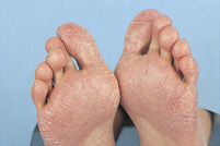
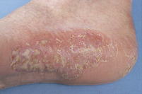
A middle-aged woman presents with a persistent, red scaly rash on the outer aspect of her foot (Fig. 26.1). As can be seen in the figure, there are yellow pustules present. She has similar lesions on the palms of her hands that have been present for a considerable time. She is a smoker.
Younger patients often also present with similar erythematous lesions. The lesion shown in Figure 26.2 occurred in a young boy who was keen on sport and frequently wore training shoes.
1 Give the diagnosis for the first condition.
2 What is responsible for the erythematous lesion in the second condition?
3 What treatment would be appropriate in both instances?
 |
| Fig. 26.2 |
 |
| Fig. 26.1 |
Plantar pustulosis/dermatosis
2 Juvenile plantar dermatosis is a scaly, glazed, fissured area of erythema seen on the weight-bearing areas of the feet, mainly across the forefeet. The skin is drier than in adult pustulosis and the condition is considered to be dermatitis from contact with synthetic materials.
3 Plantar pustulosis follows a protracted course and is often resistant to treatments such as coal tar, dithranol and steroids, although potent steroids and ultraviolet radiation may be beneficial. Treatment of the juvenile condition is easier, as often the children will be found wearing socks and shoes made from synthetic materials. They should try using an emollient cream such as Unguentum Merck and wear leather shoes if possible.
Key points
• Plantar pustulosis is a form of psoriasis seen on the feet.
• Juvenile plantar dermatosis is a contact dermatitis caused by synthetic footwear.
• Erythematous lesions are managed with steroids and emollients.
Further reading
Graham, R, Palmo-plantar pustulosis, Practitioner 233 (1989) 1428–1439;
Layton, AM; Sheehan-Dare, RA; Cunliffe, WJ, A double-blind placebo-controlled trial of topical PUVA in persistent palmoplantar pustulosis, British Journal of Dermatology 123 (s37) (1990) 44–45.
Shackelford, KE; Belsito, DV, The etiology of allergic-appearing foot dermatitis: a 5-year retrospective study, Journal of the American Academy of Dermatology 47 (5) (2002) 715–721.
Case 27
This male patient worked in the mines for many years. He is now 74 years old and regularly attends for podiatric care of his toenails. On examination, all his toenails are thickened and discoloured (Fig. 27.1). He also has macerated skin between his fourth and fifth toes and there is a red scaly patch on the side of his right foot (Fig. 27.2).


1 Why is the condition of this ex-miner’s toenails related to his previous work?
< div class='tao-gold-member'>
Only gold members can continue reading. Log In or Register to continue
Stay updated, free articles. Join our Telegram channel

Full access? Get Clinical Tree


