PROCEDURE 30 Proximal Tibia Fractures: External Fixation I
Temporary Knee Bridging External Fixation
• Immediate early fixation of complex tibial plateau fracture patterns is practiced by some surgeons. Early access to an operating room, careful preoperative planning, and meticulous surgical technique that is attentive to soft tissue handling performed by those very experienced in the management of periarticular fractures may result in good outcomes with a minimum of complications.
Indications
 Temporary knee-bridging external fixation (TKBEF) is indicated for fractures of the proximal tibia in which soft tissue injury, soft tissue swelling, or potential soft tissue swelling precludes immediate open reduction and internal fixation.
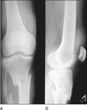
• Figure 1 shows anteroposterior (AP) (Fig. 1A) and lateral (Fig. 1B) radiographs of an extra-articular proximal tibia fracture.
 The purposes of TKBEF are
• To maintain length and alignment where there is significant comminution of the joint surface with resulting loss of stability.
• To maintain length where there is significant metaphyseal-diaphyseal comminution (i.e., Schatzker type 6 fractures).
• TKBEF may be performed in concert with surgery to restore the articular surfaces. The articular reconstruction should be done through minimal exposures to minimize the chance of wound healing problems. The use of TKBEF with this technique for articular reconstruction provides time for soft tissue swelling to dissipate prior to addressing the metaphyseal-diaphyseal component of the injury.
Examination/Imaging
 Standard AP and lateral views of the knee and proximal tibia are required.
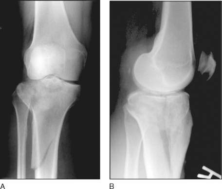
• Figure 3 shows AP (Fig. 3A) and lateral (Fig. 3B) views of a Shatzker type 6 tibial plateau fracture.
Surgical Anatomy
 The objective is to avoid introducing an iatrogenic infection into the fracture site or the regions of future surgical incisions.
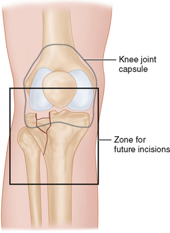
 The external fixator pins must be placed outside of the zone of injury, out of the knee joint capsule, and away from surgical incisions to be used later for definitive fixation (Fig. 5).
Stay updated, free articles. Join our Telegram channel

Full access? Get Clinical Tree


