SECTION 3 Neurology
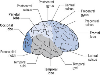
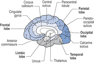
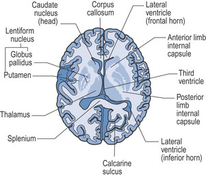
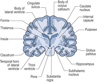
Neuroanatomy illustrations
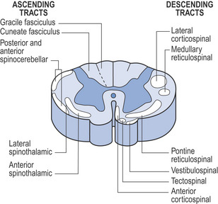
| Ascending tracts | Descending tracts |
|---|---|
| Gracile fasciculus – proprioception and discriminative touch in legs and lower trunk | Lateral corticospinal – voluntary movements |
| Medullary retrospinal – locomotion and posture | |
| Cuneate fasciculus – proprioception and discriminative touch in arms and upper trunk | Pontine reticulospinal – locomotion and posture |
| Posterior and anterior spinocerebellar – reflex and proprioception | Vestibulospinal – balance and antigravity muscles |
| Lateral spinothalamic – pain and temperature | Tectospinal – orientates head to visual stimulation |
| Anterior spinothalamic – light touch | Anterior corticospinal – voluntary movements |
Signs and symptoms of cerebrovascular lesions
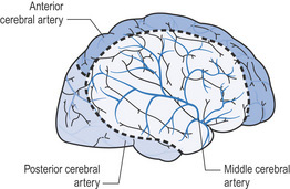
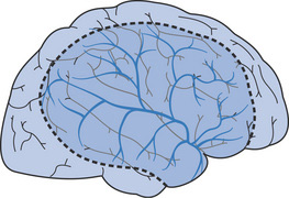
Middle cerebral artery (MCA)
| Signs and symptoms | Structures involved |
|---|---|
| Contralateral weakness/paralysis of face, arm, trunk and leg | Motor cortex (precentral gyrus) |
| Contralateral sensory impairment/loss of face, arm, trunk and leg | Somatosensory cortex (postcentral gyrus) |
| Broca’s dysphasia | Motor speech area of Broca (dominant frontal lobe) |
| Wernicke’s dysphasia | Sensory speech area of Wernicke (dominant parietal/temporal lobe) |
| Signs and symptoms | Structures involved |
|---|---|
| Neglect of contralateral side, dressing and constructional apraxia, geographical agnosia, anosognosia | Parietal lobe (non-dominant lobe) |
| Homonymous hemianopia (often upper homonymous quadrantanopia) | Optic radiation – temporal fibres |
| Ocular deviation | Frontal lobe |
| Gait disturbance | Frontal lobe (usually bilateral) |
| Pure motor hemiplegia | Posterior limb of internal capsule and adjacent corona radiata |
| Pure sensory syndrome | Ventral posterior nucleus of thalamus |
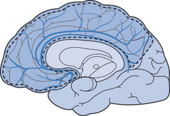
Anterior cerebral artery (ACA)
The anterior cerebral artery arises from the internal carotid artery and is connected by the anterior communicating artery. It follows the curve of the corpus callosum and supplies the medial aspect of the frontal and parietal lobes, corpus callosum, internal capsule and basal ganglia (caudate nucleus and globus pallidus).
| Signs and symptoms | Structures involved |
|---|---|
| Contralateral hemiplegia/hemiparesis (lower limb > upper limb) | Motor cortex |
| Contralateral sensory loss/impairment (lower limb > upper limb) | Somatosensory cortex |
| Urinary incontinence | Superior frontal gyrus (bilateral) |
| Contralateral grasp reflex | Frontal lobe |
| Akinetic mutism, whispering, apathy | Frontal lobe (bilateral) |
| Apraxia of left limbs | Corpus callosum |
| Tactile agnosia | Corpus callosum |
| Spastic paresis of lower limb | Bilateral motor leg area |
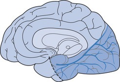
Posterior cerebral artery (PCA)
The posterior cerebral artery arises from the basilar artery. It supplies the occipital and temporal lobes, midbrain, choroid plexus, thalamus, subthalamic nucleus, optic radiation, corpus callosum and cranial nerves III and IV. The posterior communicating arteries connect the posterior cerebral arteries to the middle cerebral arteries anteriorly.
| Signs and symptoms | Structures involved |
|---|---|
| Thalamic syndrome: hemisensory loss, chorea or hemiballism, spontaneous pain and dysaesthesias | Posterior nucleus of thalamus |
| Weber’s syndrome: oculomotor paralysis and contralateral hemiplegia | Cranial nerve III and cerebral peduncle |
| Contralateral hemiballism | Subthalamic nucleus |
| Contralateral homonymous hemianopia | Primary visual cortex or optic radiation |
| Bilateral homonymous hemianopia, visual hallucinations | Bilateral occipital lobe |
| Alexia, colour anomia, impaired memory | Dominant corpus callosum (occipital lobe) |
| Memory defect, amnesia | Bilateral inferomedial portions of temporal lobe |
| Prosopagnosia | Calcarine sulcus and lingual gyrus (non-dominant occipital lobe) |
Vertebral and basilar arteries
| Signs and symptoms | Structures involved |
|---|---|
| Lateral medullary syndrome: | |
| Ipsilateral tongue paralysis and hemiatrophy | Cranial nerve XII |
| Contralateral impaired tactile sensation and proprioception | Medial lemniscus |
| Diplopia, lateral and vertical gaze palsies, pupillary abnormalities | Cranial nerve VI, medial longitudinal fasciculus |
| Bulbar palsy, tetraplegia, changes in consciousness | Bilateral corticospinal tracts |
| Pseudobulbar palsy, emotional instability | Bilateral supranuclear fibres, cranial nerves IX–XII |
| Locked-in syndrome | Bilateral medulla or pons |
| Coma, death | Brainstem |
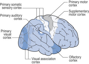
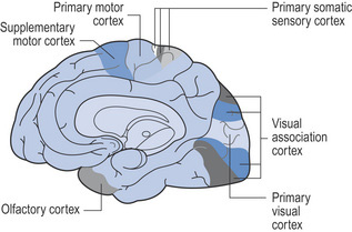
Signs and symptoms of injury to the lobes of the brain (adapted from Lindsay & Bone 2004, withpermission)
Frontal lobe
| Function | Signs of impairment |
|---|---|
| Precentral gyrus (motor cortex) | Contralateral hemiparesis/hemiplegia |
| Contralateral movement: face, arm, leg, trunk | |
| Broca/s area (dominant hemisphere) | Broca/s dysphasia (dominant) |
| Expressive centre for speech | |
| Supplementary motor area Contralateral head and eye turning | Paralysis of contralateral head and eye movement |
| Prefrontal areas ‘Personality’, initiative | Disinhibition, poor judgement, akinesia, indifference, emotional lability, gait disturbance, incontinence, primitive reflexes, e.g. grasp |
| Paracentral lobule | Incontinence of urine and faeces |
| Cortical inhibition of bladder and bowel voiding |
Parietal lobe
| Function | Signs of impairment |
|---|---|
| Postcentral gyrus (sensory cortex) Posture, touch and passive movement | Hemisensory loss/disturbance: postural, passive movement, localization of light touch, two-point discrimination, astereognosis, sensory inattention |
| Supramarginal and angular gyri | |
| Dominant hemisphere (part of Wernicke’s language area): integration of auditory and visual aspects of comprehension | Wernicke’s dysphasia |
| Non-dominant hemisphere: body image, awareness of external environment, ability to construct shapes, etc. | Left-sided inattention, denies hemiparesis |
| Anosognosia, dressing apraxia, geographical agnosia, constructional apraxia | |
| Dominant parietal lobe Calculation, using numbers | Finger agnosia, acalculia, agraphia, confusion between right and left |
| Optic radiation Visual pathways | Homonymous quadrantanopia |
Temporal lobe
| Function | Signs of impairment |
|---|---|
| Superior temporal gyrus (auditory cortex) Hearing of language (dominant hemisphere), hearing of sounds, rhythm and music (non-dominant) | Cortical deafness, difficulty hearing speech – associated with Wernicke’s dysphasia (dominant), amusia (non-dominant), auditory hallucinations |
| Middle and inferior temporal gyri Learning and memory | Disturbance of memory and learning |
| Limbic lobe Smell, emotional/affective behaviour | Olfactory hallucination, aggressive or antisocial behaviour, inability to establish new memories |
| Optic radiation Visual pathways | Upper homonymous quadrantanopia |
Stay updated, free articles. Join our Telegram channel

Full access? Get Clinical Tree


