PROCEDURE 29 Proximal Tibia Fractures: Intramedullary Nailing Max Talbot, Greg K. Berry Indications Extra-articular fractures of the proximal tibia Proximal tibia fractures with simple intra-articular extension PITFALLS • Fractures with significant tibial plateau involvement should not be treated by this method. • The proximal fragment should be large enough to accommodate three proximal locking screws. • There should be at least 4 cm of intact anterior tibial cortex in the proximal fragment to contain the nail. Treatment Options • Many nail systems are available. Choose a nail that will allow at least three proximal locking screws in different planes. • Other options Percutaneous locked plating Hybrid external fixator Open plating (not preferred) Examination/Imaging Detailed neurologic and vascular examinations, including an ankle-brachial index, are conducted. Soft tissue status and the presence of any open fractures should be determined. Compartment syndrome should be kept in mind. Anteroposterior (AP) and lateral radiographs of the entire tibia (Fig. 1A and 1B) are obtained. If there is any suspicion of articular extension, a computed tomography scan should be obtained. FIGURE 1 Surgical Anatomy 
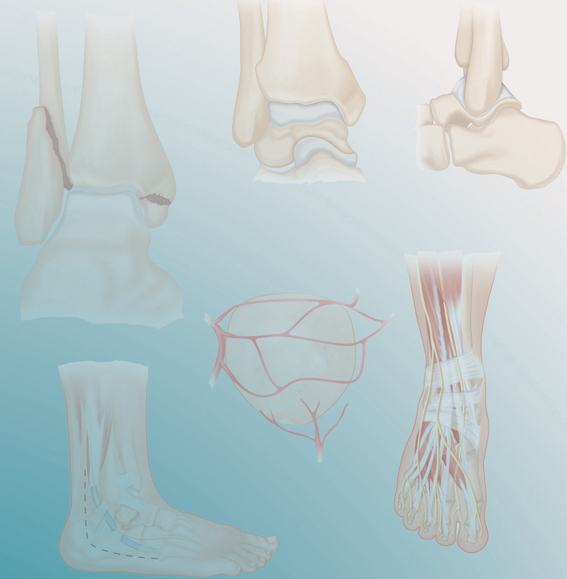
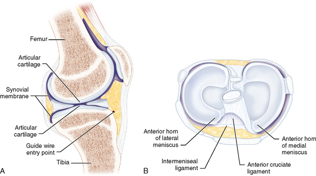
The entry point in the proximal tibia (Fig. 2A) can endanger the menisci, intermeniscal ligament, and articular surface (Fig. 2B). Use established radiologic landmarks to avoid injury to these structures. FIGURE 2 PEARLS • Prior to prepping and draping, preliminary C-arm images should be obtained to ensure that good visualization will be possible. Equipment • Adjustable triangular frame Positioning The patient is positioned supine. 
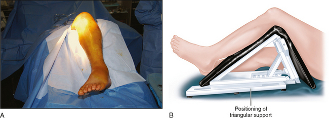
An adjustable triangular frame is used to support the limb (Fig. 3A and 3B). This allows the knee to be supported in variable degrees of flexion during the procedure. When maximal flexion is needed, we find it easier to slide the leg off the triangular frame and have an assistant hold it in the desired degree of flexion. This is mostly useful for insertion of the entry reamer, to ensure that the reamer enters the metaphysis parallel to the anterior cortex of the tibia. FIGURE 3 Portals/Exposures Either a transtendinous or a lateral parapatellar approach is recommended. Transtendinous • This is the authors’ preferred approach for proximal tibia fractures. • A skin incision is made centered on the patellar tendon, from the inferior pole of the patella to the tibial tubercle (Fig. 4). • The patellar tendon is split to access the proximal tibia. • This approach does not lead to more complications or to decreased functional outcomes compared to a parapatellar approach. Lateral parapatellar • A small incision is made lateral to the patellar tendon (see Fig. 4). • The tendon is mobilized medially. FIGURE 4 PITFALLS • With either approach, great care must be taken to protect the patellar tendon throughout the procedure, especially during reaming. PEARLS • The direction the entry reamer takes in the proximal segment is of critical importance. Only gold members can continue reading. Log In or Register to continue Share this:Click to share on Twitter (Opens in new window)Click to share on Facebook (Opens in new window) Related Related posts: 16: Perilunate Injuries 1: Open Reduction and Plate Fixation of Displaced Clavicle Fractures 7: Supracondylar Humeral Fractures 18: Femoral Neck Fractures: Arthroplasty 8: Terrible Triad Injuries of the Elbow 5: Humeral Shaft Fractures Stay updated, free articles. Join our Telegram channel Join Tags: Operative Techniques Orthopaedic Trauma Surgery Jun 7, 2016 | Posted by admin in ORTHOPEDIC | Comments Off on 29: Proximal Tibia Fractures: Intramedullary Nailing Full access? Get Clinical Tree