PROCEDURE 23 Femoral Shaft Fractures
Intramedullary Nailing
• Multiple long-bone fractures may preclude IM nailing of all fractures at a single setting due to increased risk of fat embolism.
Indications
 Reamed antegrade locked intramedullary (IM) nailing should be considered the treatment of choice for all adult femoral shaft fractures.
Examination/Imaging
• With a severe pulmonary injury or polytraumatized patient, initial external fixation followed by staged conversion to an IM nail may be indicated.
 In addition to resuscitation of the trauma patient by Advance Trauma Life Support® or similar protocol, and complete history and physical examination, focused examination of the injured extremity should include:
 Imaging should include anteroposterior (AP) and lateral plain radiographs of the femur.
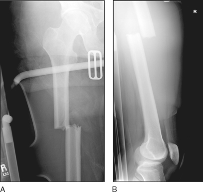
• These seldom include good-quality images of the knee and hip joints, which are essential to detect associated fractures. Figure 1 shows typical AP (Fig. 1A) and lateral (Fig. 1B) radiographs demonstrating femoral shaft fracture, but poorly visualizing hip and knee joints.
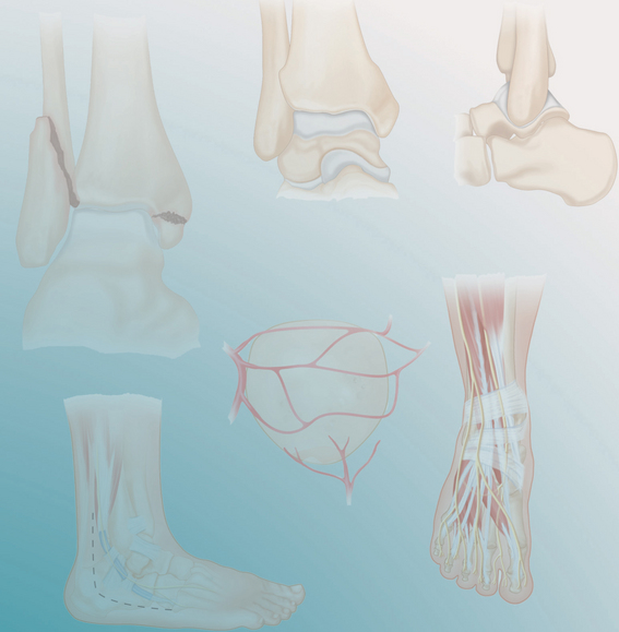
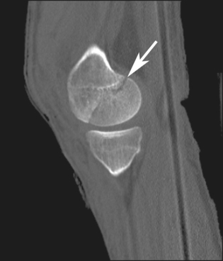
• Coronal plane (Hoffa) fractures of the distal femur (as seen in the computed tomography scan in Figure 2) and femoral neck fractures (Fig. 3) may occur with surprising frequency with high-energy femoral shaft fractures, and are easily overlooked on plain radiographs.
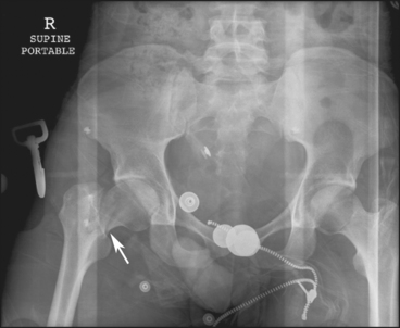
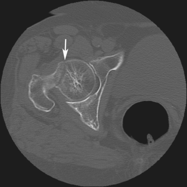
 If a computed tomography scan of the pelvis has been performed as part of the trauma assessment, look closely at the femoral neck for evidence of occult fracture (Fig. 4).
Surgical Anatomy
 The piriformis (or trochanteric) fossa lies just medial to the tip of the greater trochanter, and slightly posterior to the femoral neck, in line with the medullary canal of the femur on both AP (Fig. 5A) and lateral (Fig. 5B) views.
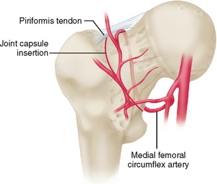
 The lateral ascending branch of the medial femoral circumflex artery runs just medial to the piriformis fossa, and its branches are at risk (Fig. 6). The piriformis and obturator internus tendon insertions are also at risk.

Positioning
• One or two surgical assistants are typically required to adequately apply traction and manipulate the fracture into a reduced position while the femur is reamed and nailed.
• Use of a fracture table to apply traction, while quite limiting in the flexibility to manipulate fracture fragments, may facilitate restoration of length and alignment without requiring skilled assistants.
 Femoral nailing is typically performed with the patient in the supine position on a flat-topped radiolucent table (Fig. 7).
 Free draping on a radiolucent table provides optimal freedom for access to, and manipulation of, the fracture for débridement (if open) and reduction.
• This position is also very useful for treatment of any associated ipsilateral lower extremity injuries (femoral neck or condyle, tibial plateau, ankle, etc.).
 A rolled flannel blanket or 3-L saline bag is placed beneath the buttock on the operative side, with the affected hip overhanging the edge of the table. Elevation of the hemipelvis on this “bump” facilitates surgical access as well as fluoroscopic imaging on the lateral view, providing a more true lateral view of the femoral neck, as well as avoiding overlap of the contralateral leg (Fig. 8).
 The leg and torso are adducted, exposing the trochanter for surgical access. The ipsilateral arm is draped over the chest to avoid interference with reaming and nailing (Fig. 9).
Stay updated, free articles. Join our Telegram channel

Full access? Get Clinical Tree


