PROCEDURE 20 Intertrochanteric Hip Fractures
Indications
 To stabilize intertrochanteric hip fractures to facilitate pain control, early mobilization, and union
 Intramedullary devices have been advocated over sliding hip screws for the treatment of more unstable intertrochanteric patterns (OTA 31-A3) (Kregor et al., 2005; Rokito et al., 1993).
• Meta-analysis comparing extramedullary devices to intramedullary devices demonstrated no significant advantages, with higher complication rates (intraoperative complications and late femoral fractures, especially with early nails) (Parker and Handoll, 2005).
Examination/Imaging
 Plain radiographs
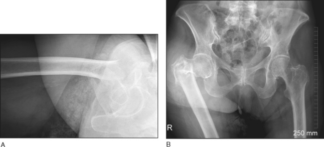
• Anteroposterior (AP) and cross-table lateral hip (Fig. 1A), AP pelvis (Fig. 1B), traction views, or full-length femur views should be obtained.
• The AP and lateral views are used to assess for signs that may suggest greater instability and potential difficulty in achieving a closed reduction or maintaining reduction:
• The AP pelvis view shows alignment of the normal side to assist in determining an adequate reduction (see Fig. 1B).
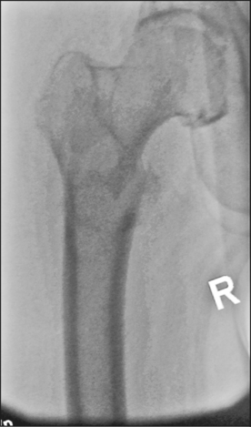
• In significantly comminuted and displaced fractures, a preoperative traction view can improve understanding of the fracture pattern, aiding in preoperative planning (Fig. 2)
Surgical Anatomy
 Gluteus medius
• The gluteus medius originates from the iliac wing inferior to the crest, inserting into the lateral and superior surfaces of the greater trochanter.
 Superior gluteal nerve
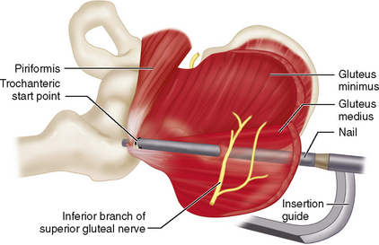
• The superior gluteal nerve arises from the lumbosacral plexus, exiting the greater sciatic notch superior to the piriformis and dividing into a superior and an inferior branch.
• The superior branch follows the line of origin of the gluteus minimus, and supplies it and the gluteus medius. The inferior branch crosses obliquely between the gluteus minimus and medius, supplying both, and terminates in the tensor fascia muscle, which it also supplies.
• The inferior limb is in close proximity to the path between the skin insertion point and the trochanteric start point, and at risk when the limb is in lower degrees of flexion and adduction (Ozsoy et al., 2007). Figure 3 shows the usual location of the nail entry in relation to the superior gluteal nerve when the hip is in the extended position.
Positioning
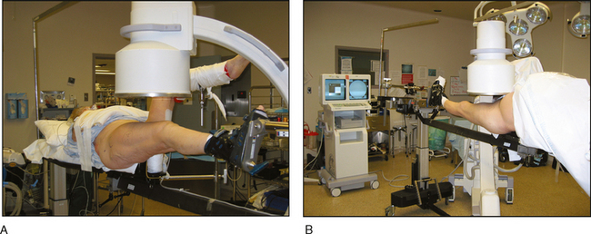
 While there is a trend toward free femoral nailing, supine positioning on a fracture table simplifies reduction and placement of the sliding screw in isolated intertrochanteric hip fractures.
• Shifting the torso to the opposite side and adducting the leg as much as possible while maintaining the reduction will make it easier to access the start point and insert the nail.
 The fluoroscopy monitor is positioned at the foot of the bed to allow both the surgeon and the fluoroscopic technician to view the image.
 Cautery and suction are also positioned at the end of the bed so they are out of the way of the C-arm.