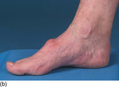
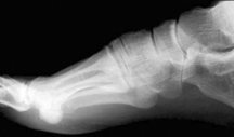
The lump on the dorsum of this middle-aged man’s foot causes him pain when he is wearing shoes (Fig. 9.1a, b). He wonders if he can have the lump removed.
1 What is the aetiology of this condition?
2 Why do the radiographs not reflect the true situation?
3 What treatment is appropriate?
Tarsometatarsal arthritis (tarsal boss)
2 The lesion is often clinically much larger than the radiographic appearance. This is due to the presence of a cartilage cap covering the bony prominence that is not evident on X-ray (Fig. 9.2).
3 Unless the condition is especially troublesome, patients are usually advised against surgery. There is a high risk of recurrence and the surgical scar may be tender where it catches any footwear. Surgical removal of only the soft tissue swelling is futile as it does not address the underlying problem. It is important to ‘saucerize’ the underlying bone and occasionally joint fusion is required.
Key points
• Minor cases can be managed with attention to footwear.
• Large bumps may cause genuine problems and surgery with excision of the underlying bone may be necessary.
• Care is required during surgery to avoid the medial dorsal cutaneous nerve.
Further reading
Parker, RG, Dorsal foot pain due to compression of the deep peroneal nerve by exostosis of the metatarsocuneiform joint, Journal of the American Podiatric Association 95 (2005) 455–458.
Case 10
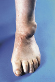
A 50-year-old man presented with a swelling on the outside of his right foot. At times it reached ‘the size of a golf ball’ (Fig. 10.1). Normally, when it reached that size he stated that his foot would become uncomfortable and he would have difficulty with shoe fitting. He would then resort to piercing the swelling with a pin, expressing a gelatinous fluid like ‘egg white’. He wondered whether he might find a more permanent cure.
1 What is the ‘egg white’ fluid that is expressed from the swelling and therefore what is the diagnosis?
2 What is the aetiopathology of this condition?
3 What should be done for this patient?
Ganglion
2 Ganglia are cystic lesions containing gelatinous fluid resulting from myxoid degeneration of connective tissue. They arise through herniation of a joint capsule or synovial sheath. They are commonly found on the dorsum of the foot because of the number of synovial sheaths passing around the ankle (Fig. 10.2). They also occur as pea-like swellings in the flexor tendon sheaths of the toes. When illuminated, these fluid-filled tumours will disperse light in contrast to opaque solid masses. The ganglion cyst has variable appearance on ultrasound but MRI demonstrates a well-defined lesion with water-equivalent signal intensity (high on T2-weighted images).
The lesion in Figure 10.3 looks similar to that shown above but in this case, the lesion was firm, adherent to the skin and non-compressible. This particular lesion did not illuminate and had low signal on T1- and T2-weighted MRI sequences. It was subsequently diagnosed as a benign fibroma but generally solid masses should be viewed with suspicion.
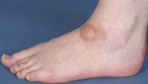 |
| Fig. 10.3 |
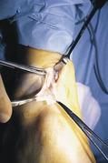
3 If asymptomatic and small, then ganglia are best left alone. Ganglia that become large or uncomfortable, as in the case illustrated above, should be aspirated to provide the patient with at least temporary relief of the symptoms. The patient should, however, be informed that the rate of recurrence from aspiration alone will be in excess of 70%. This may be reduced by corticosteroid injection although some skin discolouration and possible subcutaneous fat atrophy is to be expected. Excision of the lesion (Fig. 10.4) will provide a more certain result, provided that care is taken to ensure that the entire cyst wall is removed and any underlying bone protuberance from periosteal trauma or joint degeneration is excised.
Key Points
• Ganglia are common on the dorsum of the foot.
• Ganglia are benign lesions, containing gelatinous fluid, arising through herniation of a joint capsule or tendon sheath.
• Asymptomatic lesions do not require intervention.
• Large and uncomfortable swellings can be aspirated, but excision will often be required.
Further reading
Foo, LF; Raby, N, Tumours and tumour-like lesions of the foot and ankle, Clinical Radiology 60 (3) (2005) 308–332;


< div class='tao-gold-member'>
Only gold members can continue reading. Log In or Register to continue
Stay updated, free articles. Join our Telegram channel

Full access? Get Clinical Tree


