PROCEDURE 19 Unstable Intertrochanteric Hip Fractures
Indications for Trochanteric Stabilizing Plate
 Unstable intertrochanteric femoral fracture associated with posterolateral deficiency or lateral wall instability.
• There is a controversy over the type of the device used for an unstable intertrochanteric hip fracture.
Examination/Imaging
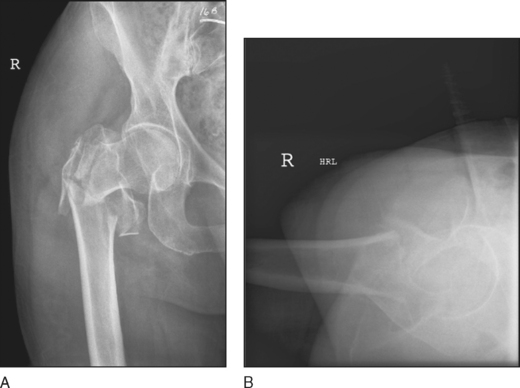
 Full femoral radiographs in anteroposterior (AP) (Fig. 1A) and lateral (Fig. 1B) views, including the joints above and below the fracture, are obtained.
 Computed tomography is less likely required (unless a femoral neck fracture is suspected and cannot be confirmed with conventional radiographs).
Surgical Anatomy
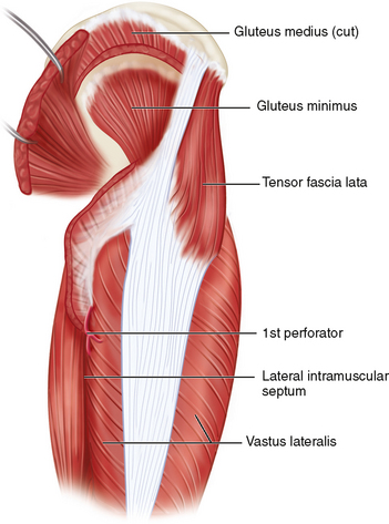
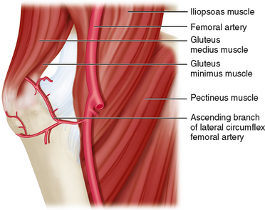
 Tensor fascia lata, glutei (medius + minimus), vastus lateralis, perforators (particularly the first perforator), and lateral intramuscular septum (Fig. 2)
• Tilting the table 10–15° to the nonoperative side might help to overcome femoral anteversion. This allows the screw direction to be parallel to the floor.
Positioning
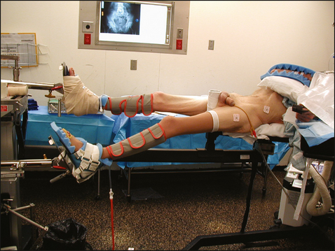
 The patient is placed in the supine position on an orthopedic fracture table, with foot-boot traction of the affected limb (Fig. 4).
• The foot is secured nicely in the boot (so it will not slip out intraoperatively when traction is applied).
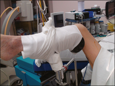
 The affected limb is kept aligned with traction, while the other limb can be placed in a scissor position, with the foot in a boot (see Fig. 4), or flexed on a Well knee-leg holder (Fig. 5).