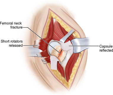
PROCEDURE 18 Femoral Neck Fractures: Arthroplasty
Indications
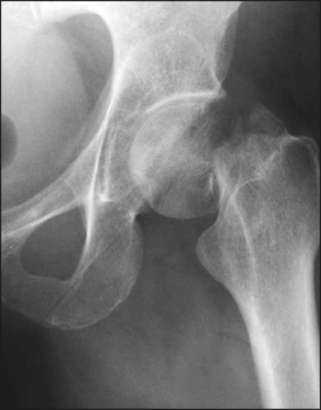
 Displaced intracapsular fractures of the femoral neck
• Absolute: elderly, osteoporotic bone, pathologic fracture, evidence of antecedent symptomatic arthritis
• Use of monopolar, bipolar, or total hip arthroplasty—differing physiologic ages, osteopenic bone, presence of acetabular degradation
• Cemented or uncemented components with a monopolar, bipolar, or total hip articulation may be used, with varying articulation sizes and materials available.
Examination/Imaging
 Occasionally computed tomography is required to elucidate complex fracture patterns and to assess posteromedial calcar.
Positioning
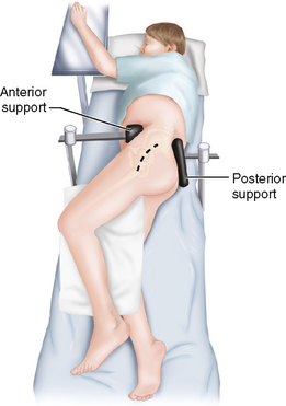
 The upper body is strapped securely with an axillary roll on the dependent side, with pressure areas supported and well padded.
Portals/Exposures
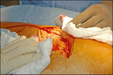
 A lateral longitudinal incision is made, centered on the greater trochanter with the proximal portion curved or angled posteriorly (Fig. 4).
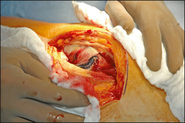
 The fascia lata is split distally in line with the femur, tensor, and gluteus maximus and proximally in line with the muscle fibers (Fig. 5).
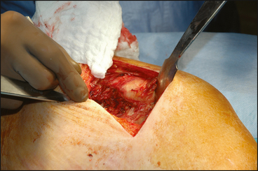
 The inferior edge of the gluteus medius is identified and retracted superiorly to expose the piriformis tendon and superior joint capsule (Fig. 6).
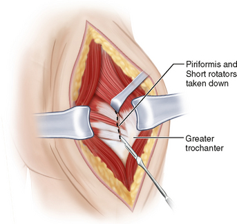
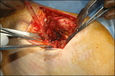
 The piriformis is released at or near its attachment deep to the posterior margin of the greater trochanter in the surgical view, and can be tagged for later identification and repair (Fig. 7).
 The remaining short external rotators are likewise released and separated from the posterior joint capsule (Fig. 8).
• A difficult internal rotation may require further release of the quadratus femoris and hip capsule toward the inferior neck.
Procedure
STEP 1: FEMORAL NECK CUT AND EXCISION OF FEMORAL HEAD
Stay updated, free articles. Join our Telegram channel

Full access? Get Clinical Tree


