PROCEDURE 11 Open Reduction and Internal Fixation of Olecranon Fractures
Introduction
 Fractures of the olecranon are common injuries and vary in scope from simple transverse fractures to complex fractures associated with injury to other structures at the elbow and forearm.


Examination/Imaging


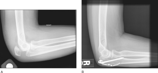
 A detailed physical examination and close inspection of plain radiographs are necessary to rule out concomitant injuries. This includes assessment of:
• Bone, joint and ligament status—the coronoid process, radial head, elbow collateral ligaments, and proximal and distal radioulnar joints


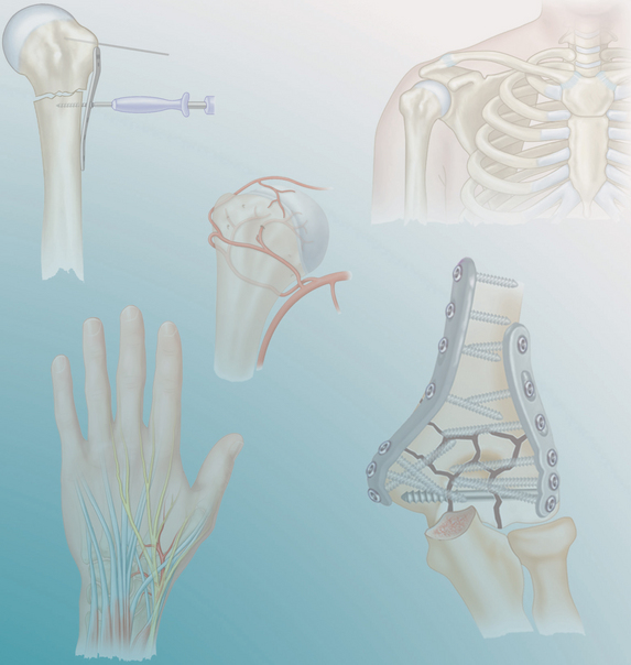
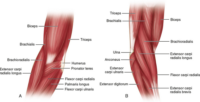
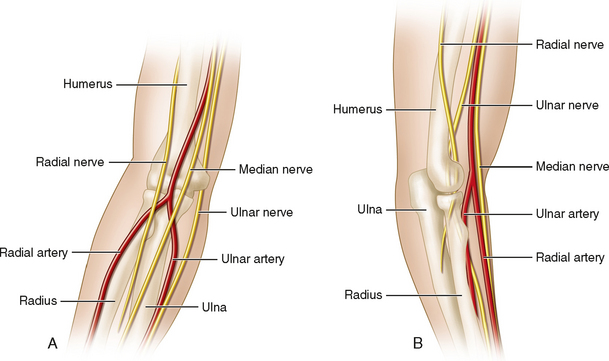
Surgical Anatomy


CLASSIFICATION AND CHOICE OF FIXATION TECHNIQUE
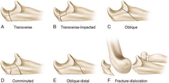
 The Schatzker classification (Fig. 4) is a simple and comprehensive scheme to describe these fractures and aids in the choice of fixation technique.
 Optimal fixation technique is dependent on fracture type.
• Type A—the classic pattern for tension band fixation (Weber and Vasey, 1963); plate and screws have also been shown to be effective.
• Type B—the joint surface impaction must be recognized, reduced, and stabilized with bone graft and/or an implant; fracture fixation as per type A.
• Type C—compression across the fracture is generated by a lag screw(s), which is protected (“neutralized”) with either plate or tension band fixation.
• Type D—reduction and fixation of the intermediate fragments with transcortical or intraosseous screws is followed by either tension band fixation (if a stable fracture configuration permitting compression is achieved) or plate and screw fixation (if the fracture configuration either does or does not permit compression).
• Type E—not mechanically amenable to tension band wiring; use plate and screws with lag technique across the fracture.