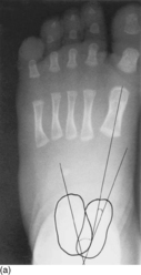
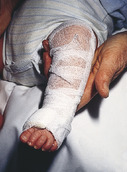
A young woman gives birth in midsummer to a daughter. It is immediately evident that the child has severe deformities of both feet (Fig. 1.1).
1 What is the approximate incidence and likely aetiology of this condition?
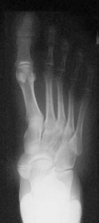
2 What are the characteristic radiological appearances?
3 What would be appropriate initial treatment?
4 At what stage would surgery be contemplated?
 |
| Fig. 1.1 |
Congenital talipes equinovarus
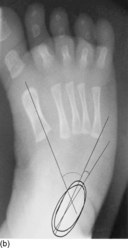
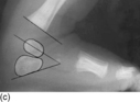
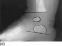
2 The primary deformities are those of hindfoot equinus and varus with forefoot adductus and supination. This positioning is secondary to medial and plantar deviation of the talar neck, medial rotation of the calcaneus and medial displacement of the navicular and cuboid. This leads to a parallel and superimposed positioning of the talus upon the calcaneus (angle <20°) and a negative talus/first metatarsal angle as shown in Figure 1.2a and b On a dorsiflexion lateral film the talocalcaneal angle is reduced (< normal 35°; Fig. 1.2c and d).
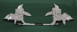
3 A gradual improvement in anatomical alignment is achieved by sequential stretching. The key, according to Ponseti, is a reversal of the cavus by dorsiflexion of the first metatarsal during the initial stage of treatment. Supination and equinus are accepted until the metatarsal is adequately dorsiflexed. Reduction in the cavus unlocks the midfoot. Subsequent correction occurs by using the uncovered talar head as a lateral fulcrum. As the forefoot is then abducted, the heel externally rotates and dorsiflexes, reversing the equinus. The position is held by applying a dynamic splint, as shown in Figure 1.3, or plaster casts applied with the knee in at least 70° of flexion. It is necessary for the physician or a trained therapist to see the child weekly to ensure that the splintage is holding the foot in the corrected position. After 2 or 3 months, if an adequate correction has been achieved, the feet may be splinted out with an abduction bar attached to the shoes. This will be used full time for 3 months and then required as a night splint to age 3 or 4 (Fig. 1.4).
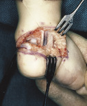
4 The optimal timing of surgery and the exact procedures required remain a matter of some debate. The majority of surgeons would probably agree that at least a lengthening of the Achilles tendon should be considered after 3 months if conservative measures fail. In Edinburgh, an extended posteromedial release through a transverse Cincinnati incision is favoured for the recalcitrant foot (Fig. 1.5), creating a mobile and plantigrade foot. The foot is immobilized in plaster following surgery to maintain the corrected position. The success of surgery relates to the compliance with bracing. Studies from the Ponseti Clubfoot Center in New York suggest that two-thirds of those non-compliant with bracing had recurrence of deformity, with one-third of these requiring more extensive surgery than Achilles tenotomy and anterior tibial tendon transfer. In contrast, only 14% of those compliant with bracing had recurrences and none required extensive surgery.
 |
| Fig. 1.3 |
 |
| Fig. 1.5 |
Key points
• Splinting, augmented by a percutaneous Achilles tendon release, will suffice in many instances.
• Surgical intervention should be considered after 3 months if conservative measures fail.
Further reading
Abdelgawad, AA; Lehman, WB; van Bosse, HJ; Scher, DM; Sala, DA, Treatment of idiopathic clubfoot using the Ponseti method: minimum 2-year follow-up, Journal of Pediatric Orthopedics 16 (2) (2007) 98–105.
Dobbs, MT; Nunley, R; Schoenecker, PL, Long-term follow-up of patients with clubfeet treated with extensive soft-tissue release, Journal of Bone and Joint Surgery 88-A (2006) 986–996.
Macnicol, MF, The management of club foot: issues for debate, Journal of Bone and Joint Surgery 85-B (2003) 167–170.
Macnicol, MF; Nadeem, RD, Evaluation of the deformity in club foot by somatosensory evoked potentials, Journal of Bone and Joint Surgery 82-B (2000) 731–735.
Ponseti, IV; Zhivkov, M; Davis, N; Sinclair, M; Dobbs, MB; Morcuende, JA, Treatment of the complex idiopathic clubfoot, Clinical Orthopaedics and Related Research 451 (2006) 171–176.
Case 2
A girl is having increasing pain when standing for prolonged periods. She is aware of tender ‘lumps’ in her insteps as shown in Figure 2.1.
1 What is causing this swelling?
2 What condition is it associated with?
3 Is surgical resection feasible?
Accessory navicular
1 This patient presents with an accessory navicular ossicle (os tibiale externum). It usually lies posteromedial to the navicular tuberosity and will almost always receive at least a part of the tibialis posterior insertion. Usually there is a small joint between the main bone and the developmental ossicle, but in some instances there is a fibrous bridge. In effect, there are two types:
• Type 1 is a small sesamoid bone within the tendon itself, anatomically separate from the navicular
• Type 2 (Fig. 2.2) is a bone developed from a separate ossification centre within the main cartilage mass of the navicular itself.
2 Patients may suffer from mild flatfoot as evident in Figure 2.1. This is rarely sufficient to prevent the patient rising onto single leg tip-toe.
 |
| Fig. 2.2 |
Key points
• The accessory navicular is a normal anatomical variant.
• 99m-Technetium bone scanning may be helpful if the significance of the accessory bone is uncertain.
• Excision of the ossicle may be necessary.
Further reading
Kopp, FJ; Marcus, RE, Clinical outcomes of surgical treatment of the symptomatic accessory navicular, Foot and Ankle International 25 (2004) 27–30.
Macnicol, MF; Voutsinas, S, Surgical treatment of the symptomatic accessory navicular, Journal of Bone and Joint Surgery 66 (1984) 218–226.
Nakayama, S; Sugimoto, K; Takakura, Y; et al., Percutaneous drilling of symptomatic accessory navicular in young athletes, American Journal of Sports Medicine 33 (2005) 531–535.
Case 3
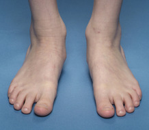
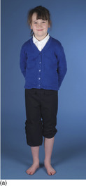
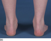
This girl’s mother is concerned about her daughter’s flat feet (Fig. 3.1a,b). She reports that her shoes develop an abnormal bulging on the medial side of the upper. There are no other symptoms and ‘her feet have always been this way’. The heel rise test is shown in Figure 3.2.
1 Describe the clinical features seen in Figure 3.1. What do these features indicate?
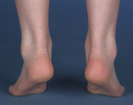
2 What is the heel rise test (Fig. 3.2), which mechanism is it testing and what does it reveal?
3 What conditions are associated with flat foot in childhood?
4 Why is this parent concerned that her daughter has flat feet?
 |
| Fig. 3.2 |
Flat foot
1 Flat foot (pes planus) is simply a low medial arch. In addition to this, the rearfoot is in a valgus alignment. In Figure 3.1 there is also evidence of Helbing’s sign (bowing of the Achilles tendon), prominence of the medial malleolus and abduction of the forefoot. The combination produces pes planovalgus.
2 For the heel rise test, patients are asked to stand on their toes. Normally this will cause the medial longitudinal arch of the foot to increase in height and the calcaneus to invert (see Fig. 7.2). With rigidity, on toe standing the arch will fail to rise and there will be no inversion of the calcaneus. This test uses Hicks’ windlass mechanism whereby dorsiflexion of the toes creates tension in the plantar fascia, drawing the forefoot and hindfoot closer. In so doing, this increases the arch and inverts the subtalar joint. An alternative to the heel rise test is Jack’s test whereby, on standing, the patient’s great toe is dorsiflexed at the MTP joint (Fig. 3.3). This should produce a similar effect to that described above. In this case, arch formation indicated that the flat foot was flexible, excluding a pathological condition such as tarsal coalition from the diagnosis (Chapter 7).
< div class='tao-gold-member'>
Only gold members can continue reading. Log In or Register to continue
Related posts:


Stay updated, free articles. Join our Telegram channel

Full access? Get Clinical Tree


