PROCEDURE 1 Open Reduction and Plate Fixation of Displaced Clavicle Fractures
Indications
• Controversy still exists regarding operative versus nonoperative treatment of clavicle fractures as nonoperative treatment has been the standard of care since the 1960s.
• Options for treatment of displaced clavicle fractures include open reduction and internal fixation or use of a sling for comfort.
• For operative treatment, open reduction and internal plate fixation is preferred as intramedullary fixation controls length and rotation poorly.
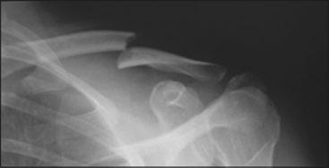
 Completely displaced midshaft clavicle fracture in young active individuals, especially with shortening
Examination/Imaging
 The length of the injured clavicle is measured from the sternoclavicular joint to the acromioclavicular joint, and compared to the opposite uninjured side both clinically and radiographically.
 A carefully documented neurovascular examination of the upper extremity is done to exclude preoperative injury.
 Anteroposterior and 20° cephalad upshot views of the clavicle are obtained to assess fracture configuration.
Surgical Anatomy
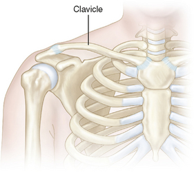
 The clavicle forms an anterior strut to maintain the position of the shoulder on the thoracic cage (Fig. 2).
• The subclavian vessels and brachial plexus pass posterior/posteroinferior to the clavicle before passing inferior to the coracoid and into the arm.
 The sternoclavicular joint is a diarthrodial joint allowing movement in both the horizontal and vertical planes, as well as 20–40° of rotation relative to the manubrium, and is stabilized by the joint capsule.
 The acromioclavicular joint is a planar joint allowing approximately 20° of rotation relative to the acromion. This joint is stabilized by the capsule and intracapsular ligaments, as well as the conoid and trapezoid coracoclavicular ligaments.
 Together, these joints allow movement of the clavicle of up to 60° in the vertical plane and 20° in the horizontal plane, and up to 40° of rotation.


