Vascular Malformations
Denise W. Metry
Mary L. Brandt
The nomenclature used to describe vascular birthmarks has long been a source of confusion in the medical literature. A biologic classification system proposed by Mulliken and Glowacki led to the current understanding of vascular birthmarks. This classification system, the most widely accepted standard for categorizing vascular birthmarks today, is the official classification system of the International Society for the Study of Vascular Anomalies. It is based on clinical behavior and cellular dynamics, and it divides vascular birthmarks into two main categories: vascular tumors (VTs) and vascular malformations (VMs).
VTs are dynamic lesions, sometimes present at birth, that are characterized by endothelial cell hyperplasia and demonstrate proliferation in infancy. In contrast, VMs almost always are present at birth (although they may not manifest until later in childhood), arise from dysmorphogenesis, exhibit normal cellular turnover, and are static or undergo slow expansion over time. VMs can be subdivided further on the basis of flow rate (fast or slow) and resemblance to vessel type (capillary, lymphatic, venous, or arteriovenous), and they can occur alone or in combination (Box 315.1). Using this classification system, more than 90% of vascular birthmarks can be distinguished from one another based on careful history and physical examination, without the need for performing ancillary studies. Accurate classification is important for understanding prognosis, potential complications, and appropriate patient management. Although most vascular anomalies are sporadic, mendelian inheritance has been observed in some families. Several causative genes, which primarily seem to affect endothelial cells, have been identified recently and have shed light on the pathophysiologic pathways involved.
BOX 315.1 Classification of Vascular Malformations
Slow-Flow
Capillary (port-wine stain)
Lymphatic
Venous
Fast-Flow
Arteriovenous
Combined (Klippel-Trenaunay Syndrome; Parkes-Weber Syndrome)
CAPILLARY MALFORMATIONS
Capillary malformations (CMs) (“port-wine stains”) are present at birth as bright red patches that may occur in any cutaneous location (Fig. 315.1). Histologically, they are composed of dilated capillary- to venule-sized vessels in the
superficial dermis, which have a reduced number of normal, surrounding nerve fibers. CMs often lighten deceptively in the first year of life, but persist thereafter. Facial CMs show a tendency to thicken, darken, and develop fibrovascular nodules, sometimes as early as during adolescence. For unknown reasons, such changes rarely occur in CMs affecting nonfacial locations. However, cutaneous CMs in any location may be associated less commonly with underlying soft tissue and skeletal hypertrophy. For example, facial CMs that involve the trigeminal V3 dermatome may result in a number of dental complications, including gingival hyperplasia, maxillary or mandibular overgrowth, and open bite deformity. Midline spinal or scalp CMs, especially those associated with other midline developmental abnormalities such as hypertrichosis, cutaneous dimples, or sinuses, can be a clue to occult spinal or cranial dysraphism.
superficial dermis, which have a reduced number of normal, surrounding nerve fibers. CMs often lighten deceptively in the first year of life, but persist thereafter. Facial CMs show a tendency to thicken, darken, and develop fibrovascular nodules, sometimes as early as during adolescence. For unknown reasons, such changes rarely occur in CMs affecting nonfacial locations. However, cutaneous CMs in any location may be associated less commonly with underlying soft tissue and skeletal hypertrophy. For example, facial CMs that involve the trigeminal V3 dermatome may result in a number of dental complications, including gingival hyperplasia, maxillary or mandibular overgrowth, and open bite deformity. Midline spinal or scalp CMs, especially those associated with other midline developmental abnormalities such as hypertrichosis, cutaneous dimples, or sinuses, can be a clue to occult spinal or cranial dysraphism.
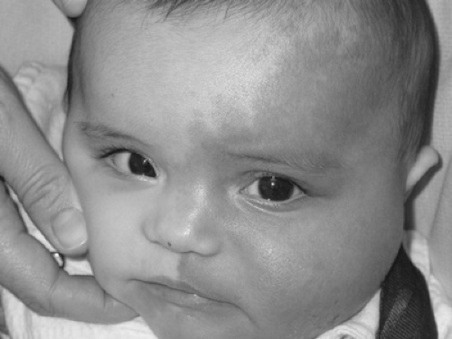 FIGURE 315.1. Capillary malformation (port-wine stain). See Color Figure 315.1 in color section. |
In most cases, CMs pose predominantly cosmetic concerns. The flashlamp-pumped, pulsed-dye laser is a well-established and safe treatment for CMs. Laser treatments may be more effective when initiated earlier in childhood, before significant vascular dilatation or hypertrophic changes occur, but other factors such as location, size, and skin pigmentation also may affect response. Individual response to therapy and the number of treatment sessions required are variable; the lateral face and trunk tend to respond better than do the extremities or central face. Treatment is safe, with minimal postprocedure discomfort and a low risk of scarring, but most CMs lighten rather than disappear completely. Because the depth of flashlamp-pumped, pulsed-dye laser penetration is less than 1.2 mm, soft tissue and skeletal hypertrophy and fibrovascular proliferation still may develop, necessitating use of other forms of intervention.
Sturge-Weber syndrome (SWS) is the association of a facial CM with ipsilateral ocular and leptomeningeal vascular anomalies. Affected children nearly always have a CM that involves the trigeminal V1 dermatome, although fewer than 10% of infants who present with this distribution have SWS. Leptomeningeal involvement, which generally can be detected by T1-weighted, gadolinium-enhanced magnetic resonance imaging (MRI), often leads to early-onset seizures, generally by the time the child reaches 2 years of age. Without prompt pharmacologic management, brain hypoxia and psychomotor deterioration may ensue. Regular ophthalmologic examinations are required for children at risk for development of SWS because increased choroidal vascularity can cause retinal detachment, glaucoma, and blindness. The Sturge-Weber Foundation is a well-established support network for affected patients and their families and for research.
Common types of vascular birthmarks that often are confused with the CM are popularly known as the “angel kiss” (forehead, eyelids, glabella, nose, and upper lip) and “stork bite” (on the nape). These birthmarks represent a minor dilatation of superficial dermal vessels, and most of them fade by the time the child is 1 to 2 years of age, although lesions of the nape often persist.
VENOUS MALFORMATIONS
VMs are structural anomalies of the venous vasculature. They generally involve the skin, subcutaneous tissues, or mucosa as soft, deep-blue masses that are easily compressible and slowly refill on release. They are present, although not always evident, at birth and can involve virtually any anatomic site. VMs swell with dependency or activity and may undergo slow expansion over time. Extensive VMs, especially those involving an extremity, can cause localized intravascular coagulopathy (LIC) owing to the chronic consumption of clotting factors, evidenced by low fibrinogen, elevated D-dimers, and a normal or moderately low platelet count. LIC also can cause the formation of phleboliths, which are a characteristic feature of VMs, even early in childhood, and are a common source of localized pain. Pain and stiffness at the site of a VM on morning awakening and dull aching are other common complaints. LIC also can lead to bleeding, which can be especially problematic when a VM involves the joint synovium.
Related posts:


Stay updated, free articles. Join our Telegram channel

Full access? Get Clinical Tree


