The purpose of this study is to investigate the failure sequence of the distal radius during a simulated fall onto an outstretched hand using cadaver forearms and high-speed X ray and video systems. This apparatus records the beginning and propagation of bony failure, ultimately resulting in distal radius or forearm fracture. The effects of 3 different wrist guard designs are investigated using this system. Serving as a proof-of-concept analysis, this study supports this imaging technique to be used in larger studies of orthopedic trauma and protective devices and specifically for distal radius fractures.
As a proof of concept, this study confirms that future studies with larger cadaveric samples can be analyzed with high-speed X ray and video to better understand distal radius fracture pathomechanics.
As a proof of concept, this study confirms that future studies with larger cadaveric samples can be analyzed with high-speed X ray and video to better understand distal radius fracture pathomechanics.
In orthopedic sports medicine and arthroplasty, dynamic fluoroscopy has been investigated to elucidate joint kinematics in vivo in attempt to understand the weight-bearing forces, shear stresses, and relative motion of the joint. To the best of the authors’ knowledge, combined high-speed X ray and video has not been used to investigate fracture pathomechanics. The objective of the current report is to serve as a proof-of-concept demonstration, evaluating the feasibility of combined high-speed x-ray and video imaging techniques applied to a fracture model. Specifically, the authors sought to investigate the fracture mechanism of the distal radius from a typical fall on an outstretched hand and the effectiveness of various wrist guard designs.
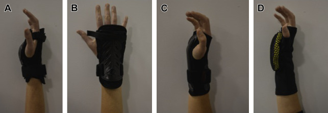
Three types of wrist guards were used, including a prototype created by the researchers. The two commercially available wrist guards were the Seirus Jam Master II (Seirus, San Diego, CA, USA), incorporating both volar and dorsal protective plates meant to be worn above a ski/snowboard glove, and the Dakine volar wrist guard (Dakine, Hood River, OR, USA), which is lower profile and consists of an aluminum volar plate. The prototype design included a viscoelastic foam core and thin rigid shell on the volar side of a neoprene sleeve ( Fig. 3 ). The appropriate size guard was secured to the cadaver hand for impact tests. The high-speed x-ray images and videos were studied for patterns of relative motion and location/sequence of fracture development. Each forearm was first tested with nondestructive loads at approximately 30% of the anticipated failure load with and without the wrist guards. Destructive tests were then performed alternating between wrist guard type and no guard.
Early results and future direction
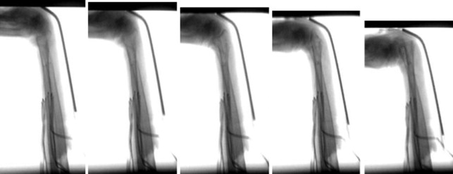
A better understanding of the pathomechanics of distal radius fractures can be gleaned by studying the videos of dynamic wrist motion when exposed to axial load, resulting eventually in fracture of the distal radius. In the authors’ study of 8 cadaver forearms, in both protected and unprotected wrists, the common failure mechanism occurred through axial compression of the extended carpus, causing the radius to shift slightly in a volar direction relative to the proximal carpal row and the proximal carpal row to hyperextend ( Figs. 4 and 5 ). The authors measured the radiocapitate angle using a goniometer to quantify the proximal carpal row hyperextension. From preload to failure the capitate extended from 42° to 55° on average. In several videos, the capitate seemed to abut the dorsal radial rim and possibly transfer the load directly to the dorsal cortex of the radius.
Related posts:

Stay updated, free articles. Join our Telegram channel

Full access? Get Clinical Tree


