Injuries to the thumb ulnar collateral ligament (UCL) are common. Failure to address the ensuing laxity of the metacarpophalangeal joint can lead to compromised grip and pinch, pain, and ultimately osteoarthritis. Instability to valgus stress with the lack of a firm end point is a strong indicator of complete rupture of the UCL. Nonoperative treatment is reserved for incomplete ruptures of the thumb UCL. Operative intervention is typically performed for complete ruptures. Repair of acute ruptures and reconstruction for chronic injuries yield excellent results. Complications are rare and most patients show preservation of motion, key pinch, and grip strength.
Key points
- •
Injuries to the thumb ulnar collateral ligament (UCL) are common. Failure to address the ensuing laxity of the metacarpophalangeal joint can lead to compromised grip and pinch, pain, and ultimately osteoarthritis.
- •
Patients who have sustained an acute injury commonly recount a traumatic episode with an accompanying onset of pain and swelling, whereas those with chronic injuries may present with less specific symptoms.
- •
The diagnosis of UCL injury relies primarily on the clinical examination of the patient.
- •
Instability to valgus stress with the lack of a firm end point is a strong indicator of complete rupture of the UCL. Ultrasonography, MRI, and plain films can be useful adjuncts.
- •
Nonoperative treatment is reserved for incomplete ruptures of the thumb UCL.
- •
Operative intervention is typically performed for complete ruptures.
- •
The goal of surgery is to restore the anatomic position of the ligament, thus providing stability to the metacarpophalangeal joint and allowing for protected range of motion early in the rehabilitation period.
- •
Both repair of acute ruptures and reconstruction for chronic injuries yield excellent results. Complications are rare and most patients show preservation of motion, key pinch, and grip strength.
- •
Range of motion exercises early in the rehabilitation phase help to ensure a positive outcome.
Introduction
Ulnar collateral ligament (UCL) injuries of the thumb are among the most common injuries in the hand. In some populations, the incidence is as high as 50 per 100,000 and they are 10 times more common than their radial version. Acute injuries, commonly referred to as skier’s thumb, have been known to occur in various sporting activities in addition to their namesake, but are also common in manual laborers. So-called gamekeeper’s thumb, the chronic form of UCL insufficiency, has also been well documented.
A stable, pain-free thumb base is not only essential for various sports and hobbies but also for activities of daily living, gripping, and key pinch. Insufficiency can lead to compromised grip and pinch, pain, and ultimately osteoarthritis. This article summarizes the current concepts in the management of these injuries in their acute and chronic forms.
Anatomy
The metacarpophalangeal joint (MCPJ) of the thumb is a diarthrodial ginglymoid (hinge) joint that has a variably flattened metacarpal head compared with its digital counterparts. Its main range of motion is in flexion and extension with a lesser amount of abduction, adduction, and rotation. The metacarpal head has a greater arc of curvature on the lateral condyle, which allows pronation with increasing flexion and enables opposition.
Stability of the MCPJ is conferred by static and dynamic stabilizers, and to a lesser amount by the articular surface. The volar (or palmar) plate with its embedded 2 sesamoid bones and the radial collateral ligament and UCL provides static stabilization. Dynamic muscular stabilizers from the intrinsics include the abductor pollicis brevis (APB), flexor pollicis brevis, and adductor pollicis (AP), as well as stabilizers from the extrinsics with the extensor pollicis longus (EPL), extensor pollicis brevis (EPB), and flexor pollicis longus (FPL). The principal dynamic stabilizer is the AP, which resists valgus forces. The FPL and intrinsic musculature overpower the EPL and EPB, producing a primary volar pull on the proximal phalanx.
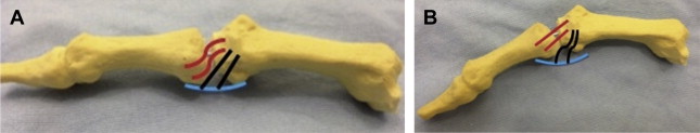
The UCL is composed of 2 distinct structures or bundles: the proper UCL and accessory UCL. The proper UCL originates from just below the metacarpal head and courses in a dorsal to palmar direction to insert on the base of the proximal phalanx. The accessory UCL lies superficial and volar to the proper UCL, blending with the volar plate and inserting on the base of the proximal phalanx. The primary function of the UCL is to provide resistance to valgus stress and volar subluxation. In extension, the accessory UCL is tight, whereas the proper UCL is lax. At approximately 30° of flexion, the proper UCL is tight, whereas the accessory UCL slides proximally with the volar plate becoming lax ( Fig. 1 ). Normal valgus laxity is approximately 6° in extension and 12° in flexion.
Introduction
Ulnar collateral ligament (UCL) injuries of the thumb are among the most common injuries in the hand. In some populations, the incidence is as high as 50 per 100,000 and they are 10 times more common than their radial version. Acute injuries, commonly referred to as skier’s thumb, have been known to occur in various sporting activities in addition to their namesake, but are also common in manual laborers. So-called gamekeeper’s thumb, the chronic form of UCL insufficiency, has also been well documented.
A stable, pain-free thumb base is not only essential for various sports and hobbies but also for activities of daily living, gripping, and key pinch. Insufficiency can lead to compromised grip and pinch, pain, and ultimately osteoarthritis. This article summarizes the current concepts in the management of these injuries in their acute and chronic forms.
Anatomy
The metacarpophalangeal joint (MCPJ) of the thumb is a diarthrodial ginglymoid (hinge) joint that has a variably flattened metacarpal head compared with its digital counterparts. Its main range of motion is in flexion and extension with a lesser amount of abduction, adduction, and rotation. The metacarpal head has a greater arc of curvature on the lateral condyle, which allows pronation with increasing flexion and enables opposition.
Stability of the MCPJ is conferred by static and dynamic stabilizers, and to a lesser amount by the articular surface. The volar (or palmar) plate with its embedded 2 sesamoid bones and the radial collateral ligament and UCL provides static stabilization. Dynamic muscular stabilizers from the intrinsics include the abductor pollicis brevis (APB), flexor pollicis brevis, and adductor pollicis (AP), as well as stabilizers from the extrinsics with the extensor pollicis longus (EPL), extensor pollicis brevis (EPB), and flexor pollicis longus (FPL). The principal dynamic stabilizer is the AP, which resists valgus forces. The FPL and intrinsic musculature overpower the EPL and EPB, producing a primary volar pull on the proximal phalanx.
The UCL is composed of 2 distinct structures or bundles: the proper UCL and accessory UCL. The proper UCL originates from just below the metacarpal head and courses in a dorsal to palmar direction to insert on the base of the proximal phalanx. The accessory UCL lies superficial and volar to the proper UCL, blending with the volar plate and inserting on the base of the proximal phalanx. The primary function of the UCL is to provide resistance to valgus stress and volar subluxation. In extension, the accessory UCL is tight, whereas the proper UCL is lax. At approximately 30° of flexion, the proper UCL is tight, whereas the accessory UCL slides proximally with the volar plate becoming lax ( Fig. 1 ). Normal valgus laxity is approximately 6° in extension and 12° in flexion.
Pathoanatomy
Injury to the UCL usually occurs as the result of hyperabduction or hyperextension of the MCPJ. This injury is commonly found among a high number of sports and falls onto an outstretched hand. The term skier’s thumb was coined for its high incidence among skiers who fell, with the pole causing injury to the thumb. Tearing of the UCL also commonly results in concomitant injury to the dorsal capsule and the volar plate leading to a volar subluxation of the proximal phalanx. This subluxation is caused by the palmar/radial force of the APB causing a radial shift. Because the UCL travels dorsal to the flexion-extension axis, chronic UCL insufficiency can lead to a supination deformity at the proximal phalanx as it rotates around an intact radial collateral ligament. Each of these deformities can be evident on clinical examination or radiographic assessment.
The UCL tear most commonly occurs at its distal insertion, sometimes as an avulsion fracture, but midstance and proximal ruptures do occur. If the abduction force continues to pull the distal phalanx radially, the adductor aponeurosis can travel far enough distal to cause the UCL to lie superficial to the adductor aponeurosis. This condition is termed a Stener lesion and precludes the UCL from healing by nonoperative means. This Stener lesion has been noted to occur in approximately 60% to 90% of cases, depending on the source.
Physical examination
After obtaining a thorough history, examination begins with inspection of the thumb. In acute injuries, swelling and ecchymosis are usually evident and volar radial subluxation may be visible with larger injuries. Pain is elicited on the ulnar aspect of the MCPJ with palpation. In some cases, a palpable mass may be evident, suggesting a Stener lesion.
Stress examination of the MCPJ is the most important evaluation of stability of the UCL and should be compared with the contralateral side. One hand should stabilize the metacarpal neck while the other grasps the proximal phalanx and controls rotation, because rotation of the phalanx can masquerade as valgus laxity ( Fig. 2 ). Valgus stress should be applied to the thumb in full extension and in 30° of flexion. Laxity of either 30° total or 15° more than the contralateral side suggests injury. If the laxity occurs in 30° of flexion alone, an injury to the proper UCL is suspected. If instability is noted in 30° of flexion and also in full extension, injury to the proper and accessory UCL is suspected. If instability is noted only in extension, the volar plate may be the only injured structure. Malik and colleagues showed that a difference of 15° in flexion or extension can be normal in some patients, but the numbers stated earlier are traditionally accepted. Because of this variation, some clinicians prefer to place more emphasis on whether or not there is a firm end point to stress testing, which differentiates a partial from a complete injury.
Stressing the MCPJ in an acutely injured patient may be painful, with guarding or muscle spasm giving a false-negative examination. Local anesthesia can be injected into the MCPJ to allow for a better examination, which has been shown to increase accuracy from 28% to 98% in a study of 47 patients.
Diagnostic studies
Radiographs should always be obtained for patients with a history and physical examination suggesting a UCL tear. Radiographic assessment can identify avulsion fractures, isolated fractures of the proximal phalanx, or associated fractures of the metacarpal. Radial and volar subluxation can also be seen, as well as a supination deformity of the proximal phalanx relative to the metacarpal, termed the sag sign ( Fig. 3 ). Stress radiographs may be performed to better assess the degree of laxity in those patients with an equivocal clinical examination ( Fig. 4 ). Some investigators suggest radiographic assessment before stressing the UCL because stress risks displacement of fracture fragments; however, if the initial energy was not strong enough to displace the fragment, then intraoffice stress is not likely to be adequate to change its position. Fractures, which can occur in up to 40% of injuries, are generally of 2 different patterns, as described by Hintermann and colleagues. One is a true avulsion fracture and the other is an isolated fracture that is not associated with tear of the UCL. Differentiation can be made with stress radiographs. In a true avulsion fracture, stress in the radial direction causes further displacement of the fracture from the ulnar corner of the proximal phalanx.
Ultrasonography (US) is a noninvasive and cost-effective modality to evaluate for UCL tear when used along with clinical examination ( Fig. 5 ). Results have varied widely, with accuracy ranging from 40% to 92%. Review of the literature on US examination of UCL tears shows an overall sensitivity of 76%, specificity of 81%, accuracy of 81%, positive predictive value of 74%, and a negative predictive value of 87%. Variability exists because the diagnostic value of US largely depends on the experience of the performing individual.
MRI is less cost-effective but more consistent in its ability to aid in the diagnosis of UCL tears, especially with dedicated extremity coils ( Fig. 6 ). Arthrography has been shown to be even more accurate than MRI alone. Hergan and colleagues showed sensitivity and specificity of 100% with MRI versus 88% and 83% respectively with US.
It is the author’s opinion that advanced imaging with US or MRI should be obtained in cases in which history or clinical examination is uncertain. US may be more cost-effective if an experienced musculoskeletal radiologist is available to perform the examination; if not, MRI should be obtained.
Management
The question of nonoperative or operative treatment is largely in debate but the decision depends on stability and associated fractures. UCL injuries are generally categorized in grades. Grade I injuries, also termed sprains, are tender along the UCL but without increased laxity. Grade II injuries have increased laxity with a firm end point and are thought to be partial tears. Grade III injuries have both increased laxity and no firm end point, which indicates a complete tear. Grade I and II injuries are commonly treated nonoperatively. Complete ruptures, with or without associated Stener lesions, are treated surgically without consensus on optimal timing. Both repair and reconstruction have shown satisfactory results with regard to pain, strength, motion, and stability. Unstable avulsion fractures are treated as grade III ligament injuries and isolated displaced fractures are treated with reduction and fixation of the articular segment.
Nonoperative treatment
Grade I and II injuries are appropriate for nonoperative treatment, because they are considered stable injuries. For these injuries with pain, mild laxity, and firm end points, immobilization is required to allow resolution of inflammation and to protect the MCPJ from further injury during the healing phase. The type of immobilization varies by clinician, but appropriate means include short-arm thumb spica casting, hand-based thumb spica casting, custom hand-based thermoplast splints, removable thumb spica splints, and functional hinged splints. The important aspect is to protect the MCPJ from stress while allowing the interphalangeal (IP) joint of the thumb to move freely, thus avoiding undue stiffness.
Length of immobilization ranges from 10 days to 6 weeks depending on the extent of injury (sprain vs partial tear); however, immobilization for a period of 4 weeks is typically recommended. Athletic participation is generally acceptable if the form of immobilization is permitted by local sporting regulations and use of the thumb is not required for that position or sport. At 4 weeks, occupational therapy or physiotherapy is initiated to regain motion, particularly in the flexion/extension plane, while avoiding valgus stresses at the MCPJ. At this time, immobilization is only required in high-risk activities, whether on the playing field or in the work or recreational environment. Strengthening begins at 6 to 8 weeks depending on the patient’s progress, with unrestricted activity usually permitted at 12 weeks.
Nonoperative treatment has been reported in grade III injuries and UCL avulsion fractures with good results. Landsman and colleagues and Pichora and colleagues reported stable pain-free thumbs in 85% and 90% of their grade III injuries, respectively. However, the remaining patients in each study continued to have pain and instability, eventually requiring operative intervention. Pursuing nonoperative treatment of grade III injuries should be done cautiously and appropriate patient education should be performed at the outset.
Nonoperative treatment of UCL avulsion injuries has also shown mixed results in the literature. If there is a firm end point with a nondisplaced or minimally displaced fragment, immobilization alone can be considered. Kuz and colleagues and Sorene and Goodwin showed that, despite a 20% and 60% fibrous union rate respectively, all their nonoperatively treated patients were satisfied with their outcomes. This finding is in contrast with reports by Dinowitz and colleagues and Bowers and Hurst who reported persistent pain and instability after immobilization in all or nearly all of their patients, with most requiring eventual operative treatment. Dinowitz and colleagues showed that triangular-shaped articular avulsion fragments may undergo rotation whereby the articular surface rotates into the fracture gap, whereas plain radiographs often appear to show a simple nondisplaced fracture. Close scrutiny is required to avoid mischaracterization of these seemingly nondisplaced fractures that are certain to lead to nonunion and likely posttraumatic arthritis, pain, and stiffness.
Related posts:

Stay updated, free articles. Join our Telegram channel

Full access? Get Clinical Tree


