Tibial stress fractures are common in the athlete. There are various causes of these fractures, the most common being a sudden increase in training intensity. Most of these injuries are treated conservatively; however, some may require operative intervention. Intervention is mostly dictated by location of the fracture and failure of conservative treatment. There are several surgical options available to the treating surgeon, each with advantages and disadvantages. The physician must understand the nature of the fracture and the likelihood for it to heal in a timely manner in order to best treat these fractures in this patient subset.
Key points
- •
Tibial stress fractures are common in the athlete. There are various causes of these fractures, the most common being a sudden increase in training intensity.
- •
Most of these injuries are treated conservatively; however, some may require operative intervention. This is mostly dictated by location of the fracture, as well as failure of conservative treatment.
- •
There are several surgical options available to the treating surgeon, each with advantages and disadvantages.
- •
The physician must understand the nature of the fracture and the likelihood for it to heal in a timely manner in order to best treat these fractures in this patient subset.
Tibial stress fractures are relatively common in athletes, with a reported incidence as high as 10% to 20% in runners. Treatment varies based on a number of factors, including cause, location, and duration of symptoms. Most tibial stress fractures heal uneventfully with conservative treatment; however, some can be more challenging, ultimately requiring surgery. In athletes, time to recovery is a strong consideration in the decision algorithm. Missing competition for an extended period of time can have a psychosocial impact, and, in the case of professional athletes, an economic impact. Physicians must know which fractures will likely heal in a reasonable time period and which fractures are prone to delayed union or nonunion.
Tibial stress fractures are caused by repetitive microtrauma that results in increased osteoclastic activity and an imbalance between resorption and regeneration of bone. They commonly occur in individuals participating in strenuous activity such as military training and athletics. Certain sports such as running have a high occurrence of stress fractures, with the tibia being the most common site.
Underlying intrinsic and extrinsic factors can predispose individuals to developing these fractures. Intrinsic factors include hormonal imbalances, nutritional deficiencies, metabolic bone diseases, relative avascularity of an area of bone, and muscle imbalances. Extrinsic factors include sudden increases in the intensity of an exercise regime. Often the intrinsic and extrinsic causes are related and may occur simultaneously. The female athletic triad of amenorrhea, disordered eating, and low bone mineral density results from an excessive training regimen coupled with decreased caloric intake. Low body fat percentage leads to a decrease in estrogen levels with resultant osteoporosis and a propensity toward stress fracture.
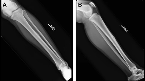
Stress fractures commonly occur in 3 areas of the tibia: posteromedial cortex, anterior cortex, and tibial plateau. The posteromedial cortex is the most common site. Stress fractures in the anterior cortex are less common, with an incidence of around 4% of all tibial stress fractures. The anterior tibial cortex is the tension side of the bone, and stress fractures in this area can be more problematic, as the healing potential is lower. The dreaded black line has been described as a V or wedge-shaped defect in the anterior cortex ( Fig. 1 ). They tend to be more common in athletes who participate in jumping and leaping activities, such as gymnasts and basketball players, but they also occur in runners.
History
Appropriate treatment depends on prompt and accurate diagnosis. Patients report insidious onset of pain without a history of trauma. Pain is worsened by activity, particularly repetitive loading. Initially, pain increases at the end of activity and may be relieved with rest. Gradually, symptoms worsen and may persist beyond activity, frequently at night. Patients often describe an acute change in training regimen with an increase in activity.
The physician should elicit a complete history related to diet, menstrual function, athletic background, and training regimen. Dietary factors to consider extend beyond disordered eating. Low calcium intake and insufficient vitamin D have been associated with lower extremity stress fractures. Low-fat diets also have been shown to increase risk. Low energy availability and excessive exercise, in addition to disordered eating, alter menstrual function and lead to low bone mineral density (BMD).
Females with low body-mass index, amenorrhea, and low mean muscle mass are at increased risk of stress fracture. One or more components of the female athletic triad—amenorrhea, disordered eating, and low BMD—often are present. Of the three, menstrual dysfunction has been implicated as the most important.
Gymnasts, cross country runners, and dancers are at increased risk as well. Attention should be paid to both volume and intensity of workouts. Abrupt changes or increases in training often precipitate a stress fracture. Physicians should recognize conditions associated with osteoporosis or osteopenia including vitamin deficiencies, endocrinopathies, alcoholism, and renal dysfunction.
Physical examination
Tenderness typically is localized with palpation over a diaphyseal stress fracture. Percussion away from the fracture often causes pain. Specific examination tests have been described and can be useful. The hop test, single leg hopping, and the fulcrum test, performed by placing a bending moment on the tibia against either the end of the examination table or the physician’s knee or arm, produce severe localized pain in patients with tibial stress fractures. Swelling and overlying skin changes such as bruising should be noted. Possible causative factors should be examined. Structural malalignment, leg length discrepancy, foot deformities, low lean muscle mass, and smaller calf girth can predispose to increased tibial stress.
Differential diagnosis
Stress fractures are the result of normal bone being subjected to abnormal stress. By contrast, insufficiency fractures refer to normal stress applied to abnormal, pathologic, or osteoporotic bone. Pathologic fractures should always be considered, paying particular attention to any history of cancer and other constitutional systemic symptoms. Significant pain without fracture may be present with an osteoid osteoma or other bone tumors.
Medial tibial stress syndrome (MTSS) or traction periostitis (shin splints) causes medial leg pain that usually resolves promptly with conservative treatment. Exertional compartment syndrome and nerve entrapment also should be excluded. A high index of suspicion for infection, both superficial and osteomyelitis, should be maintained, particularly in children and adolescents. Common conditions such as sprains, strains, tendonitis, contusions, and muscle soreness must be excluded. Finally, stress reactions, which are prefractures, present with progressive symptoms.
Among the differential diagnosis for tibial stress fractures, distinguishing posteromedial stress fractures from medial tibial stress syndrome can be challenging. The 2 entities are sometimes grouped together in the literature as part of a continuum of stress reactions. Distinctions have been made with regard to etiology and pathology. MTSS is theorized to be a traction-induced periositis of the medial tibia combined with repetitive bending loads across the tibia. The soleus, which originates from the medial aspect of the tibia along with the flexor digitorum longus, acts as an inverter of the foot and contracts eccentrically during midstance as the foot pronates. Studies have demonstrated that athletes who have increased pronation during midstance are more at risk for MTSS.
MTSS is diagnosed based on history and physical examination. Clinically, the distal two-thirds of the posteromedial border of the tibia are diffusely tender to touch, and soft tissue swelling may be present. Advanced imaging should be used only when the diagnosis is in question or when conservative treatment fails to relieve symptoms. On MRI, MTSS appears as diffuse periosteal edema. Bone scanning has proven to be sensitive in diagnosing MTSS, with a longitudinal area of uptake present on the delayed phase of a triple-phase scintigraphy corresponding to the diffuse area of periostitis. In contrast, all 3 phases of the scan are positive in acute stress fractures.
Imaging
Plain radiographs often are the first imaging modality used; however, radiographs have a low sensitivity (10% to 50%) for detecting stress fractures, particularly early in the clinical course. Predictable progression in findings includes pretibial swelling, thickening of the cortex, and finally a visible fracture on plain films. If initial films are negative, repeat imaging in 2 weeks is indicated. Anteroposterior, lateral, and oblique films should be taken.
MRI has proven to be the best imaging modality for diagnosing fractures that are not visible on plain films, as it is both highly sensitive and specific ( Figs. 2 and 3 ). One study comparing MRI with bone scan and computed tomography (CT) showed the sensitivity and specificity of MRI to be 82% and 100%, respectively. Increased signal in the endosteum on fat-suppressed T2-weighted images occurs shortly after the onset of symptoms, prior to evidence on plain radiographs. MRI also provides bony and soft tissue detail that can help differentiate stress fractures from other pathology. This ability can prove especially important in distinguishing tibial stress fractures from MTSS. More severe fractures will have a distinct fracture line apparent on both T1-and T2-weighted sequences.
Related posts:

Stay updated, free articles. Join our Telegram channel

Full access? Get Clinical Tree


