The Forearm
Pronator Teres
Patient Position:
Supine with the forearm supinated.
Needle Insertion:
Place the index finger on the cubital fossa and insert the needle medially to the finger at approximately 5 centimeters (cm) below the elbow crease.
Activation:
Pronate the forearm with the elbow slightly flexed.
Clinical Notes:
If inserted too deeply, the needle may penetrate the median nerve or brachial artery; too medially, the flexor carpi radialis.
Innervation:
C6,C7-upper and middle trunk-anterior division-lateral cord-median nerve.
Origin:
Medial epicondyle of the humerus and coronoid process of the ulnar.
Insertion:
Lateral surface of the radius to the midline (wraps around the radius).
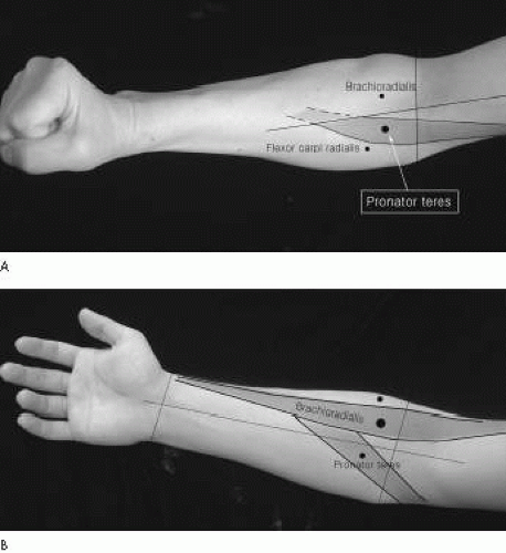 Figure 12-1. Pronator teres. |
Flexor Carpi Radialis
Patient Position:
Supine with forearm supinated.
Needle Insertion:
Insert the needle at the proximal one-third to one-half between the tendon of flexor carpi radialis at the wrist and the medial supracondylar area of humerus. It is medial to the pronator teres. This muscle is medial to the pronator teres. It is a superficial muscle and is not clearly demarcated from the adjacent muscles.
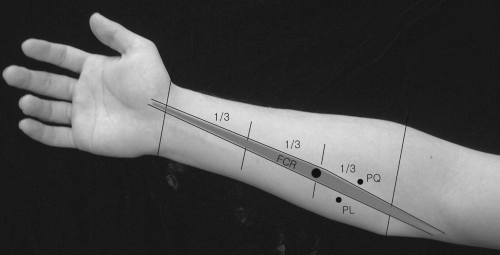 Figure 12-2. Flexor carpi radialus. |
Activation:
Flex and abduct the wrist (radial deviation).
Clinical Notes:
This muscle may be involved in pronator syndrome. If the needle is inserted too laterally, it will be in the pronator teres; if too medially, it will be either in the palmaris longus; if too deeply, it will be in the flexor digitorum superficialis or flexor digitorum profundus.
Innervation:
C6,C7-median nerve.
Origin:
Medial epicondyle of the humerus by a common flexor tendon.
Insertion:
Base of the volar surface of the second metacarpal bone.
Palmaris Longus
Patient Position:
Supine with forearm fully supinated.
Needle Insertion:
Insert the needle at the proximal one-third of a line drawn between the palmaris longus tendon at the wrist crease and medial epicondyle.
Activation:
Flex the wrist.
Clinical Notes:
The muscle is superficial, slender, fusiform, and medial to the flexor carpi radialis. If inserted too deeply, it will be in the flexor digitorum
superficialis. This muscle is absent in 10% to 13% of the population and is subject to a lot of variation.
superficialis. This muscle is absent in 10% to 13% of the population and is subject to a lot of variation.
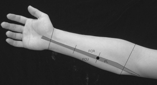 Figure 12-3. Palmaris longus. |
Innervation:
C7,C8-median nerve.
Origin:
Medial epicondyle of the humerus by a common tendon.
Insertion:
Palmar aponeurosis and flexor retinaculum.
Flexor Digitorum Superficialis (Sublimis)
Patient Position:
Supine with forearm supinated.
Needle Insertion:
Identify the tendons of palmaris longus and flexor carpi ulnaris at the wrist and follow proximally to the midforearm where the needle is inserted. The muscle is more superficial at this point.
Activation:
Flex the middle phalanx of each of the medial four digits.
Clinical Notes:
If the needle is inserted too deeply, it will be in the flexor digitorum profundus. Entrapment of the anterior interosseous nerve occurs by either a fibrous origin of this muscle or a tendinous origin of it to the third digit.
 Figure 12-4. Flexor digitorum superficialis (sublimis). |
Innervation:
C7,C8 and T1-median nerve.
Origin:
Medial epicondyle of the humerus by a common flexor tendon, coronoid process of the ulna, and ulnar collateral ligament of the elbow joint.
Insertion:
Palmar aspect of the middle phalanx of the medial four digits.
Flexor Pollicis Longus
Patient Position:
Supine with forearm supinated.
Needle Insertion:
Insert the needle at the junction of middle and distal third of the forearm between the brachioradialis and flexor carpi radialis. Before inserting, note the radial pulse at the insertion site.
Activation:
Flex the distal phalanx of the thumb.
Clinical Notes:
In diagnostic workup of carpal tunnel syndrome, this muscle is examined to rule out possible more proximal involvement of the median nerve.
Innervation:
C8, T1-lower trunk-medial cord-anterior interosseous branch (nerve) of the median nerve.
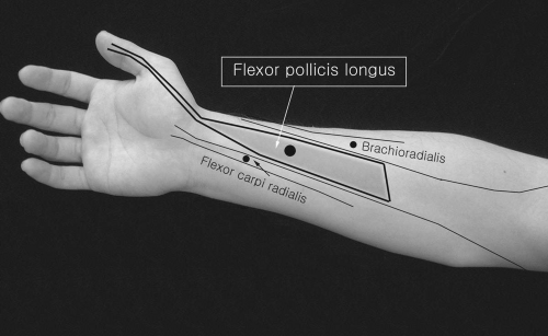 Figure 12-5. Flexor pollicis longus. |
Origin:
Anterior surface of the middle half of the radius, adjoining the anterior interosseous membrane.
Insertion:
Base of the volar surface of the distal phalanx of the thumb.
Pronator Quadratus
Patient Position:
Supine with forearm supinated or pronated.
Needle Insertion
Related posts:


Stay updated, free articles. Join our Telegram channel

Full access? Get Clinical Tree


