The Anterior Thigh
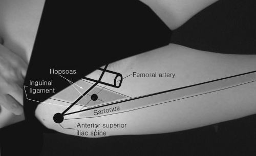
Iliopsoas
Patient Position:
Supine with the hip slightly flexed and the knee in a comfortable position.
Needle Insertion:
Activation:
Flex, adduct, and externally rotate the thigh.
Clinical Notes:
The lumbar plexus is located within this muscle and the femoral artery descends through the psoas major and then runs between it and the iliacus. If the needle is inserted too laterally, it will reach the sartorius.
Innervation:
The iliacus is the most proximal muscle innervated by the femoral nerve (L2, L3), whereas the psoas major is innervated by ventral rami of the first, second, and third lumbar spinal nerves.
Origin:
Psoas major from bodies and transverse processes of the 12th thoracic and all the lumbar vertebrae; iliacus from the superior aspect of the iliac fossa.
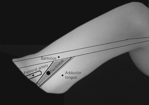
Pectineus
Patient Position:
Supine.
Needle Insertion:
Palpate the femoral artery pulse in the inguinal area and insert the needle about 1 centimeter (cm) medial to the artery and below the inguinal fossa. If inserted too deeply, the needle will be in the obturator externus: too medially, in the gracilis or adductor longus.
Activation:
Ask the patient to flex and adduct the hip.
Clinical Notes:
Flat, quadrilateral-shaped muscle occasionally innervated by the obturator or accessory obturator nerve.
Innervation:
L2, L3-femoral nerve.
Origin:
Pectineal line of the pubis.
Rectus Femoris
Patient Position:
Supine with the knee extended.
Needle Insertion:
Insert the needle at the middle third of the femur anteriorly and halfway between the medial and lateral border of the thigh.
Activation:
Flex the hip with the knee extended.
Clinical Notes:
Flat, spindle-shaped muscle. It is the only “quadriceps” muscle that crosses two joints. If the needle is inserted too deeply, it will be in the vastus intermedius.
Innervation:
L2, L3, L4-femoral nerve.
Origin:
Anterior inferior iliac spine, upper lip of acetabulum, and fibrous capsule of the hip joint.
Insertion:
Base of the patella and by the patella ligament into the tibial tuberosity.
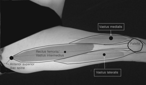 Figure 17-3. Rectus femoris. |
Vastus Medialis
Related posts:


Stay updated, free articles. Join our Telegram channel

Full access? Get Clinical Tree


