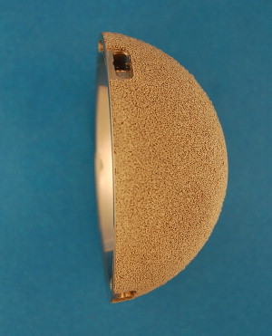
Resurfacing systems use press-fit, monoblock, cobalt chrome alloy acetabular sockets because of the material’s ability to withstand stresses while accommodating a large femoral head. Despite the widespread use of these types of sockets for both hip resurfacing and total hip replacement, there is a paucity of literature assessing the outcomes of these cups in particular. The 10 year survivorship of the Conserve® Plus monoblock acetabular component used in this study was 98.3% with small pelvic osteolytic lesions suspected in only 2.3%. This study highlights the excellent radiographic survivorship profile of the Conserve® Plus socket.
Reports of the durability of certain first-generation metal-on-metal total hip arthroplasty implants led to a renewed interest in this bearing couple in the 1980s. Low wear rates associated with metal-bearing surfaces and the material’s ability to withstand stresses have enabled larger head sizes required for resurfacing arthroplasty of the hip. Large-diameter femoral heads continue to be attractive for total hip arthroplasty because they provide increased stability and decreased dislocation rates. Previously, large diameter heads in metal-on-polyethylene articulations were impractical, given the unacceptable high amount of volumetric wear. Over the past decade, many hip surgeons have become interested in metal-on-metal resurfacing arthroplasty for end-stage arthrosis in young active patients.
Resurfacing systems typically use press-fit, monoblock, cobalt chrome alloy acetabular sockets. These sockets differ from the modular titanium shells, which are typically used in conventional total hip replacement. Monoblock cobalt chrome cups are stiffer than their titanium counterparts and they do not allow for initial screw fixation and thus are entirely dependent on stability from a sound press-fit at initial implantation. Furthermore, concerns have arisen because of heightened awareness of the potential for adverse local soft tissue reaction (ALTR) to metal-on-metal bearings, particularly with certain designs.
Outcomes data on the longevity of these sockets are needed. The purpose of this study was to define the midterm survivorship and radiographic results of a cobalt chrome alloy monoblock acetabular component in patients who undergo hip resurfacing.
Materials and methods
A retrospective review was performed, which included the first 643 hips in 580 consecutive patients who were treated with hip resurfacing arthroplasty using the Conserve® Plus prosthesis (Wright Medical Technology, Arlington, TN, USA) between November 1996 and October 2003. The surgical technique used for implantation of the prostheses has been previously described All hips were implanted with the original acetabular component, which has a constant thickness of 5 mm. The Conserve® Plus acetabular shell is made of cobalt, chromium, and molybdenum, with a porous coating of sintered beads 75 to 150 μm thick for cementless fixation ( Fig. 1 ). The mean age of the patients at the time of surgery was 48.9 years (range, 14–78 years) and 75% were men. Their mean weight at the time of surgery was 83.5 kg (range, 42–164 kg) and body mass index (BMI, calculated as the weight in kilograms divided by height in meters squared) 27 (range 17–46). The causes were distributed as follows: osteoarthritis 66%, developmental dysplasia of the hip 10%, osteonecrosis 8%, trauma 8%, inflammatory diseases 3%, developmental or metabolic diseases 4%, other 1%. The patients were followed up for 4 months after surgery and then yearly. Whenever the patient could not come to the center for follow-up visits, radiographs performed in another institution were requested and a phone follow-up consultation scheduled. The senior author (H.C.A) also performed outside clinics in 20 different locations throughout the United States to reach those living outside Southern California. The radiographic review was performed by 2 experimenters (J.B.H and S.T.B) blinded to the clinical results of the patients. All immediate postoperative radiographs were analyzed for acetabular component position, including abduction angle, anteversion, presence of gaps between the acetabular cup and the reamed bone, and percent bone coverage in the frontal plane. Abduction and anteversion of the cup were measured using Ein-Bild-Röentgen-Analysis cup software, version 2003 and the contact patch to rim (CPR) distance computed from these data. From the last follow-up films and with comparison to the immediate postoperative films, the presence and size of radiolucencies in the DeLee-Charnley zones were recorded. Evidence of cup migration, pelvic osteolysis, and stress remodeling of the periacetabular bone of the pelvis were also noted. Heterotopic ossification was recorded using the grading system of Brooker and colleagues. Kaplan-Meier survival estimates were calculated using 2 different end points:
- 1.
Revision for aseptic loss of acetabular component fixation.
- 2.
Radiographic loss of acetabular fixation defined by a complete radiolucency covering all 3 De Lee-Charnley zones.

Radiolucency was used for further analysis, and Cox proportional hazard ratios were computed to identify possible risk factors specific to the durability of the acetabular component.
Results
The average duration of the follow-up period was 10.4 years (range 7.1–14.0 years), and the mean radiographic follow of the series was 6.8 years (range 0.1–13.4 years). From this cohort, 482 hips had a radiographic follow-up collected during the fifth year after surgery or beyond. In this series, 45 hips underwent revision surgery at a mean time to revision of 61.6 months (range 1–139 months). However, the reason for revision was a loss of acetabular component fixation in only 5 hips revised at a mean of 75.2 months (range 2–120 months): At 5, 8, 9 and 10 years, 4 acetabular components loosened, and 1 component protruded through the medial acetabular wall 4 days after surgery. Of these hips, 3 were revised to a conventional THR, whereas 2 were maintained as hip resurfacings by insertion of a new cup of greater outside diameter ( Fig. 2 ).
The Kaplan-Meier survival estimate of the acetabular component was 99.6% at 5 years (95% confidence interval, 98.6%–99.9%) and 98.3% at 10 years (95% confidence interval, 95.5%–99.4%) when revision for loss of acetabular fixation was used as end point. In addition, 5 hips showed complete radiolucencies covering the 3 De Lee-Charnley zones around the cup at a mean follow-up time of 115.9 months (range, 94–127 months). Using the time to radiographic failure of these hips and the hips revised for acetabular loss of fixation as end point ( Fig. 3 ), the Kaplan-Meier survival estimate of the acetabular component was 99.6% at 5 years (95% confidence interval, 98.6%–99.9%) and 97.6% at 10 years (95% confidence interval, 94.6%–98.9%).
The mean cup abduction angle was 43.5° (range 16°–72°). The mean anteversion angle of the cup was 17.4° (range 2°–51°). The mean CPR distance was 14.5 mm (range 1–25 mm), and in 79 hips, the CPR distance was less than 10 mm. Acetabular cup coverage was complete in 60.8% of the hips, ranging from 70% to 100%. All hips included, radiolucencies were visible in zone 1 for 19 cups (including 2 radiolucencies 2 mm thick), in zone 2 for 22 cups (including 4 radiolucencies 2 mm thick), and in zone 3 for 36 cups (including 6 radiolucencies 2 mm thick). Radiolucencies in multiple zones were seen in 15 cups (2.3%), including 9 cups (4 revised) with radiolucencies in all 3 zones (which were included in the second survivorship analysis). Small amounts of cup migration (≤2 mm) were observed in 7 cups (1.0%). Small pelvic osteolytic lesions were suspected in 15 hips (2.3%). Brooker grade III heterotopic ossification was seen in 25 hips (3.9%) and Brooker grade IV in 6 hips (0.9%).
Postoperative gaps were observed in 30 cups (4.7%) in zone 1 (4 of 2 mm or more), 118 cups (18.4%) in zone 2 (35 of ≥2 mm), and 7 cups (1.1%) in zone 3 (2 of ≥2 mm). All postoperative gaps filled in within 36 months, except in 8 hips; 4 cups settled into the original gap and appeared stable, whereas 3 showed a fibrous fixation at first follow-up. One cup was revised at 36 months for sepsis. There was no association of the presence of a gap on the postoperative radiograph and the occurrence of cup aseptic loosening.
Young age (hazard ratio 0.942, P = .022), low BMI (hazard ratio 0.816, P = .027), and low CPR distance (hazard ratio 0.848, P = .037) were identified as risk factors for a higher rate of cup failure to osseointegrate or subsequent loss of fixation.
Results
The average duration of the follow-up period was 10.4 years (range 7.1–14.0 years), and the mean radiographic follow of the series was 6.8 years (range 0.1–13.4 years). From this cohort, 482 hips had a radiographic follow-up collected during the fifth year after surgery or beyond. In this series, 45 hips underwent revision surgery at a mean time to revision of 61.6 months (range 1–139 months). However, the reason for revision was a loss of acetabular component fixation in only 5 hips revised at a mean of 75.2 months (range 2–120 months): At 5, 8, 9 and 10 years, 4 acetabular components loosened, and 1 component protruded through the medial acetabular wall 4 days after surgery. Of these hips, 3 were revised to a conventional THR, whereas 2 were maintained as hip resurfacings by insertion of a new cup of greater outside diameter ( Fig. 2 ).


