Imaging
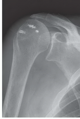
Any preoperative evaluation for possible revision rotator cuff repair should start with plain radiographs. While further imaging studies are likely to be needed, basic radiographs may direct the surgeon’s choice of subsequent studies or immediately rule out revision rotator cuff repair as a viable option all together, such as in patients with bone changes suspicious for chronic osteomyolitis, an arthritic glenohumeral joint, or patients with fixed superior migration of the humeral head (
Fig. 27-1).
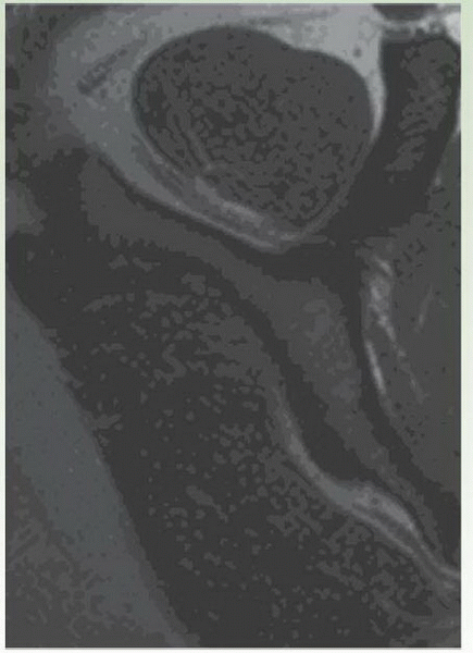
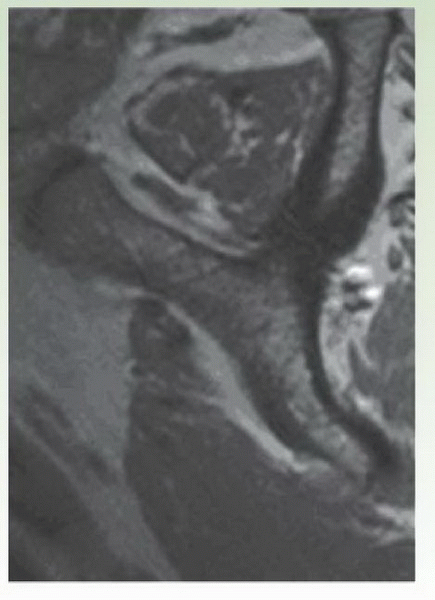
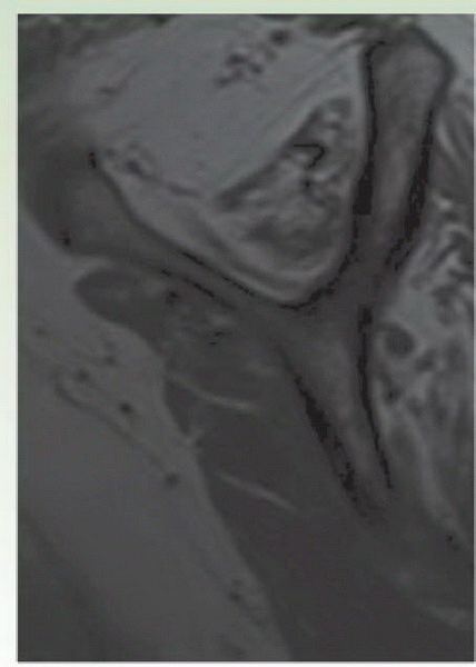
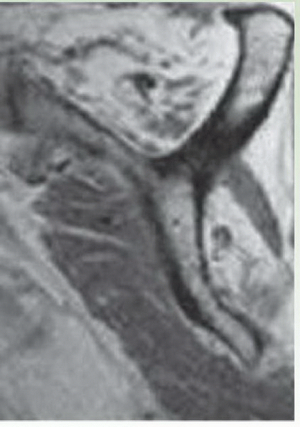
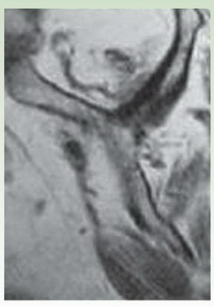
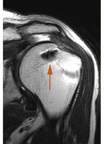
MRI is the most commonly obtained follow-up imaging study to evaluate the shoulder girdle when considering revision surgery. Concern is often given to the ability to adequately visualize the rotator cuff when an MRI is performed in the setting of retained metallic suture anchors in the humeral head. While metal artifact often obscures the bone immediately adjacent to the anchor(s), it has not been our experience that retained metal anchors in the humeral head significantly compromise the ability to evaluate the essential characteristics of a failed rotator cuff repair, and for that reason, this is still our imaging modality of choice (
Fig. 27-2). The tear location should be assessed to help the surgeon to develop a surgical strategy (open vs. arthroscopic approach; likelihood of the need for soft-tissue releases; fixation strategies; etc.) and correlated with physical exam findings. Greater degrees of rotator cuff muscle degeneration have been demonstrated to correlate with lower structural healing rates and worse functional outcomes following surgery (
1,
2); therefore, these characteristics should be specifically and carefully evaluated on MRI in order to accurately estimate the chances for success if revision repair is to be undertaken. The magnitude of medial tendon retraction seen on MRI should also be assessed. Although it has not been shown to correlate directly with repair success, a retracted tendon is more likely to require extensive soft-tissue releases to mobilize the tendon prior to repair and is more likely to result in a repair under higher tension, which is believed to be one of the primary causes of postoperative repair failure (
11).
It is important to note that superior migration of the humeral and the acromiohumeral distance are not characteristics well evaluated using MRI or CT scan since superior translation of the humeral head may occur in patients when they lie down and gravity is no longer pulling the humeral head inferiorly. Such dynamic superior migration is not known to be associated with lower rotator cuff repair success rates and is distinctly different from static or “fixed” superior migration of the humeral head, which is most effectively identified on plain radiographs and will be present even when the patient is standing/sitting.
Computed tomography (CT) arthrogram is another commonly used imaging modality to evaluate the size and location of a rotator cuff tear when planning revision surgery. In addition to being faster to obtain and less expensive, it is also an easy alternative for people who are unable to obtain MRIs for other reasons (claustrophobia, retained intracranial clips, cardiac pacemakers, etc.). This modality is very effective for determining the size, location, and tendon retraction associated with a rotator cuff tear. However, it can
suffer from problems with metal artifact, and additionally, several studies have suggested that it is more difficult to assess degenerative muscle changes with this modality than with MRI (
12). Although rotator cuff tendon integrity can be assessed with this imaging method, some attributes of the tendon such as the presence of tendonosis, intratendonous delamination, and tendon thickness can be difficult if not impossible to assess.
Ultrasound is another less expensive imaging option for evaluating rotator cuff tears that has become the study of choice for some institutions. Performed by a skilled ultrasonographer, this modality can be used to determine tear size, location, the magnitude of tendon retraction, and even the degree of rotator cuff muscle degeneration and intrasubstance changes (tendonosis) within the tendon itself. The greatest limiting factor to use of this modality is that it is highly user dependent. Detailed and accurate information about the tear characteristics requires skilled and experienced group of people performing the study and interpreting the findings. Most people experienced with using this modality emphasize the importance of evaluating dynamic imaging— previously recorded video or preferably real-time images as opposed to static pictures—in order to accurately assess the tear. Body habitus (very muscular or morbidly obese patients) or limited motion of the shoulder can also make adequate imaging of the rotator cuff difficult if not impossible with this modality.
Laboratory Studies
In addition to the standard lab work needed for general preoperative medical clearance, a C-reactive protein and erythrocyte sedimentation rate as well as white blood cell count should be obtained to rule out occult infection on any patient planned for revision rotator cuff repair. Patients with known diabetes should also obtain a hemoglobin A1C to confirm that their blood glucose level is well controlled. For those patients with a recent history of smoking, consideration should be given to checking the blood for a nicotine level so that the true smoking status of the patient at the time of surgery is known.
