Stress Fractures of the Foot
Selene Parekh
Cameron Ledford
CLINICAL PRESENTATION
Stress fractures are overuse injuries most commonly seen in athletes and military recruits; however, the incidence is rising in the general population secondary to earlier and longer participation in recreational activity. Stress fractures are most common in the weight-bearing bones of the lower extremity most commonly noted in the tibia, tarsals, and metatarsals. These injuries result from repetitive, submaximal loading of a bone leading to microfracture. These breaks are unable to heal secondary to imbalances between bone resorption and formation propagating into a fully identifiable fracture. Patients typically present with a progressive onset of pain over a period of days to weeks. They usually experience an increase in symptoms with activity and a relief with rest. There may not be a specific inciting event, but often symptoms are associated with a change in activity level or training regimen. A thorough history including diet, nutrition, medications, daily activities, footwear, menstrual cycles in females, and sleep patterns should be discussed.
CLINICAL POINTS
Patients report progressive pain especially after increased intensity or duration of activity.
Lower extremity bones are most common including tibia, tarsals, and metatarsals.
Most common in athletes, military recruits, and females with menstrual irregularities
PHYSICAL FINDINGS
On physical exam, point tenderness is almost universal and often will identify the bone involved. The pain may be diffuse initially but will then localize to the site of the fracture. Pain with range of motion of a joint around a stress fracture may also be elicited. In more superficial areas, edema, warmth, ecchymosis or even a palpable callus may be present. Assessment of limb alignment and length discrepancies, gait, passive range of motion, tendon function, and calluses provide the examiner information about repetitive stresses put on the symptomatic area.
STUDIES (LABS, X-RAYS)
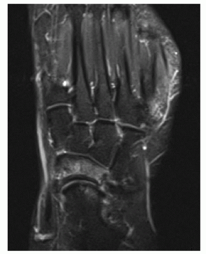
Imaging studies including radiographs, computed tomography (CT) scans, MRI, and bone scintigraphy can be helpful, especially when the diagnosis is questionable or is suspected in a high-risk bone. Multiple views of weight-bearing plain films would include an anteroposterior, lateral, and oblique (Fig. 33-1). These images will often be negative for the first 2 weeks following a stress fracture, until callus formation
occurs. Eventually, a fracture line, callus, lucency, or sclerosis may be visible. Radionuclide bone scan has been shown to be a sensitive imaging modality, and uptake in all three phases of a technetium-99 m diphosphonate scan can be seen in the first 48 to 72 hours (see Fig. 33-2). MRI has replaced bone scan as the imaging modality of choice in many settings and gives superior specificity and resolution (see Fig. 33-3). Cortical defects and bone marrow edema are best visualized on T2-weighted images and suggest the presence of stress fracture. In addition, MRI imaging is helpful in grading the stage of stress fractures and, therefore, can more accurately predict the time to recovery. CT scan can be used to identify incomplete and complete fractures but cannot aid in identification of stress reactions (see Fig. 33-4). Therefore, CT scan is thought to be more helpful than MRI in following the healing of stress fractures.
occurs. Eventually, a fracture line, callus, lucency, or sclerosis may be visible. Radionuclide bone scan has been shown to be a sensitive imaging modality, and uptake in all three phases of a technetium-99 m diphosphonate scan can be seen in the first 48 to 72 hours (see Fig. 33-2). MRI has replaced bone scan as the imaging modality of choice in many settings and gives superior specificity and resolution (see Fig. 33-3). Cortical defects and bone marrow edema are best visualized on T2-weighted images and suggest the presence of stress fracture. In addition, MRI imaging is helpful in grading the stage of stress fractures and, therefore, can more accurately predict the time to recovery. CT scan can be used to identify incomplete and complete fractures but cannot aid in identification of stress reactions (see Fig. 33-4). Therefore, CT scan is thought to be more helpful than MRI in following the healing of stress fractures.
Stay updated, free articles. Join our Telegram channel

Full access? Get Clinical Tree


