Stepwise Approach to Uniplane, Biplane, Delta, and Hybrid External Fixation
Paul S. Cooper
Introduction
Uniplanar (uniplane) or monolateral (unilateral) external fixators are devices applied to one side of the lower extremity commonly affixed with half-pins. Due to the limits of half-pin fixation, these external fixation devices are viewed for short-term use, when compares with that of transosseous wire multiplanar versions. Indications for monolateral external fixation include temporary stabilization in acute trauma settings, limb lengthening, ankle arthrodesis and arthrodiastasis. Additional applications include midfoot and hindfoot arthrodesis and as an adjunct for wound healing through soft tissue stabilization. While technically simpler to apply, these external fixation devices are limited in the scope of deformity correction. Hybrid fixation combines advantages of transosseous wires distally and half pin fixation proximally.
Uniplane Monolateral Ankle External Fixator Technique
With the patient in slight external rotation position, a monolateral external fixation device is chosen that has sufficient length to span the ankle joint and secure half-pins in the tibia above the level of pathology (Figures 7.1 and 7.2). The first half-pin inserted is dictated by the talar half-pin, which is directed into the talar neck perpendicular to the medial talus (Figure 7.3). This ideal entry point is located about one finger’s breadth anterior to that of the medial malleolus and advanced to obtain bicortical fixation. Once this half-pin is confirmed to be in the optimal location, the ankle clamp of the monolateral external fixator is slid over the half-pin. This serves as a half-pin template for placement of the second half-pin in the medial calcaneus is directed (Figure 7.4). The second half-pin is drilled through the medial calcaneal body with bicortical fixation. Soft tissue considerations of this half-pin insertion include the calcaneal nerve superficially and the contents of the tarsal tunnel deeper. Careful exposure consists of first creating a dimple corresponding to the half-pin site insertion. Longitudinal stab wound followed by dissection to bone with a small hemostat minimizes the incidence of a calcaneal nerve injury (Figures 7.5–7.7). The ankle construct is then secured and tightened. The ankle hinge is left free, as well as the hinges on the proximal and distal ends of the monolateral fixator. In addition, the compression–distraction segment is set at the half-point of excursion. With all mobile segments free, the upper tibial half-pins are directed medial or anteromedial at maximum pin spread (Figures 7.8 and 7.9). Once these half-pins are secured on the tibia, the ankle is positioned in the desired plane with regard to dorsiflexion and plantarflexion, rotation and varus/valgus alignment, and the articulated hinges are locked both proximally and distally (Figures 7.10–7.12). The final adjustment comes at the ankle hinge where the final degree of ankle sagittal plane position is adjusted. This position may be unlocked by the patient postoperatively for ankle active range of motion if desired (Figures 7.13–7.16 and Clinical Case I). Following this, the compression–distraction device is attached and the central unit is unlocked as needed for axial adjustment (Figures 7.17 and 7.18).
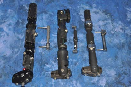 Figure 7.1. Examples of uniplane monolateral ankle external fixators. Note each has a compression/distraction central unit. |
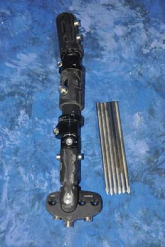 Figure 7.2. Uniplane monolateral ankle external fixator setup for application includes the pre-built unit and self-drilling half-pins. |
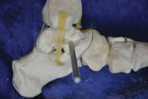 Figure 7.3. First half-pin is inserted into the talar neck in perpendicular orientation to the ankle joint. |
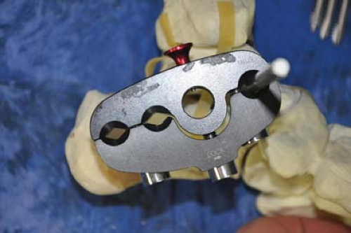 Figure 7.4. The pin clamp is applied and used for additional half-pin insertion template. |
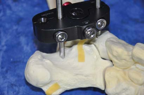 Figure 7.5. Second half-pin aligned over the calcaneal body. This approximates the location of the neurovascular bundle and initial dissection to bone will minimize neurovascular complications. |
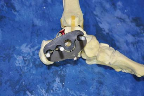 Figure 7.6. Optional third pin inserted into the calcaneal body. Note orientation of clamp in relation to the talus and calcaneus. |
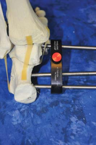 Figure 7.7. Dorsal view demonstrating half-pin positioning. |
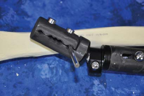 Figure 7.8. Proximal half-pin inserted into medial tibia. Note upper clamp is fully unlocked on ball hinge to allow freedom for optimal positioning. |
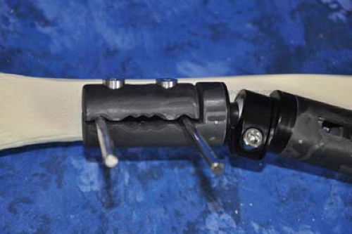 Figure 7.9. Second half-pin inserted at the farthest proximal hole in the tibia to obtain maximum stability. Additional half-pins may be added as needed. |
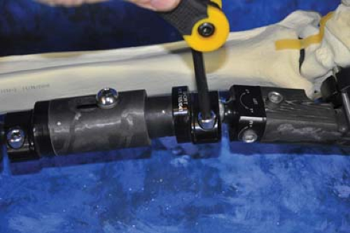 Figure 7.10. Securing the distal hinge first. The ankle should be positioned in neutral dorsiflexion/plantarflexion before tightening. |
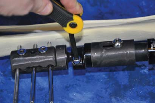 Figure 7.11. The proximal clamp hinge is then tightened. |
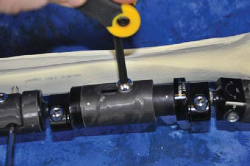 Figure 7.12. The central compression/distraction unit is locked. This can be released later once the unit is applied. |
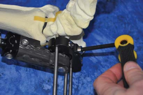 Figure 7.13. Final ankle sagittal plane position is locked into the ankle hinge joint. |
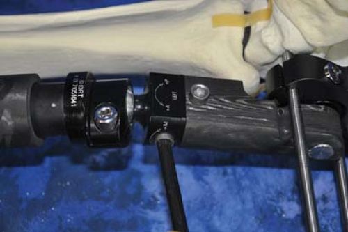 Figure 7.14. Some uniplane monolateral ankle external fixators allow for residual correction in other planes. In this example, translation of the joint can be adjusted. |
 Figure 7.15. Final anterior view of the static uniplane monolateral ankle external fixator. |
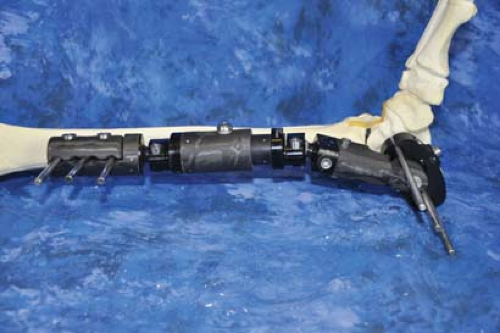 Figure 7.16. Final medial view of the static uniplane monolateral ankle external fixator. |
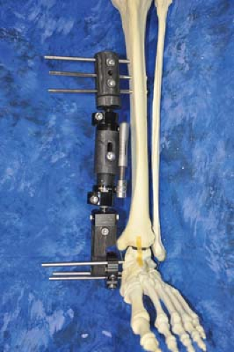 Figure 7.17. Final construct with compression/distraction unit applied. |
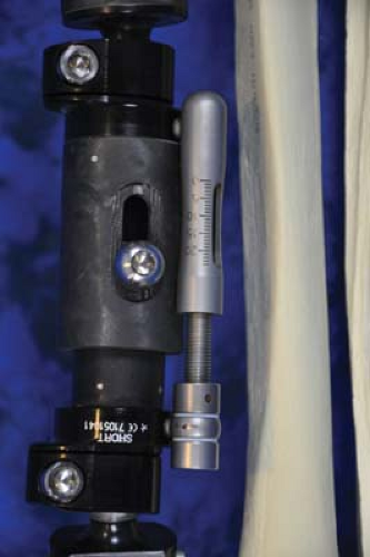 Figure 7.18. Close-up of compression/distraction unit used in this specific uniplane monolateral ankle external fixator to allow for axial adjustments. |

Biplane Ankle External Fixator Technique
With the patient positioned in a supine position, the basic setup of the biplane external fixator is chosen (Figures 7.19–7.21). Next, a four-hole pin fixation clamp is chosen for the proximal tibia. The clamp should have extensions for bar to bar clamps on both sides. The first pin is inserted perpendicular to the tibial crest either with self-drilling half pins, or conventional methods of stab wound, soft tissue dissection and half-pin insertion (Figures 7.22–7.24). The pin clamp is then positioned two finger’s breadth from the anterior tibia and lightly tightened, to be used as a guide for subsequent pin insertions. The next pin is inserted parallel with the tibial crest using the farthest hole to maintain optimal pin spread (Figures 7.25–7.28). Additional pins may be inserted between these two pins if additional fixation deemed necessary (Figures 7.29 and 7.30).
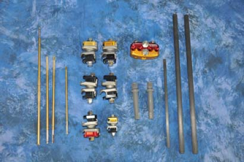 Figure 7.19. Basic setup for construction of the biplane ankle external fixator consisting of four-hole clamp, two pin clamp extension bars, four bar to bar large clamps, one bar to bar small clamp, small bar, and half-pins. |
Bars that span the ankle with sufficient length are chosen for medial and lateral connections (Figure 7.31). Distally, a transfixion pin is drilled through the hindfoot in the calcaneal body directed medial to lateral perpendicular to the longitudinal axis of the foot (Figures 7.32–7.34). A small incision with dissection down to the bone minimizes potential injury
to the medial calcaneal nerve. The transfixion calcaneal pin is inserted to optimally cover all threads under the subcutaneous tissue if possible. Following this, medial (Figure 7.35) and lateral (Figure 7.36) extension bars are placed off the tibia pin clamp. Proper length connecting bars are chosen and attached to the tibial fixation block and that of the calcaneal transfixion pin (Figures 7.37 and 7.38). The ankle is positioned in neutral dorsiflexion, but will fall into equinus with only a single transfixion pin into the hindfoot (Figures 7.39 and 7.40 and Clinical Case II).
to the medial calcaneal nerve. The transfixion calcaneal pin is inserted to optimally cover all threads under the subcutaneous tissue if possible. Following this, medial (Figure 7.35) and lateral (Figure 7.36) extension bars are placed off the tibia pin clamp. Proper length connecting bars are chosen and attached to the tibial fixation block and that of the calcaneal transfixion pin (Figures 7.37 and 7.38). The ankle is positioned in neutral dorsiflexion, but will fall into equinus with only a single transfixion pin into the hindfoot (Figures 7.39 and 7.40 and Clinical Case II).
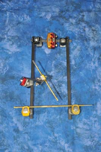 Figure 7.20. Biplane ankle external fixator used for acute lower extremity trauma. Note the half-pin extension for the forefoot and the threaded transfixion pin for the calcaneus.
Related posts: History and Evolution of External Fixation History and Evolution of External Fixation
 Stepwise Approach to Static Circular External Fixation Stepwise Approach to Static Circular External Fixation
 Stepwise Approach to Forefoot Elective and Reconstructive Surgery with External Fixation Stepwise Approach to Forefoot Elective and Reconstructive Surgery with External Fixation
 Stepwise Approach to Ankle Elective and Reconstructive Surgery with External Fixation Stepwise Approach to Ankle Elective and Reconstructive Surgery with External Fixation
 Surgical Off-loading External Fixation and Plastic Reconstruction of the Foot and Ankle Surgical Off-loading External Fixation and Plastic Reconstruction of the Foot and Ankle
 Stepwise Approach to Bone Loss, Transport, and Lengthening with External Fixation Stepwise Approach to Bone Loss, Transport, and Lengthening with External Fixation
Stay updated, free articles. Join our Telegram channel
Full access? Get Clinical Tree
 Get Clinical Tree app for offline access

|