Stepwise Approach to Midfoot/Hindfoot Trauma and External Fixation
John J. Stapleton
Vasilios D. Polyzois
Thomas Zgonis
Introduction
The utilization of external fixation for midfoot and hindfoot trauma has increased due to the successful outcomes of external fixation in treating certain ankle and pilon fractures. One of the most common indications for external fixation is the management of selected open and/or high-energy midfoot/hindfoot trauma. The focus of this chapter is to discuss in a detailed fashion the application of various external fixation designs for the treatment of midfoot and hindfoot fractures and/or dislocations.
Indications/Contraindications
Severe midfoot crush injuries usually present with significant bone loss and pulverized fracture patterns that involve the medial column (navicular and/or medial cuneiform), the lateral column (cuboid and/or anterior process of the calcaneus), and/or the central column (navicular and intermediate/lateral cuneiforms). External fixation is utilized to stabilize and reestablish these columns and to obtain anatomic alignment if definitive open reduction and internal fixation (ORIF) cannot be achieved. In addition, open fractures of the midfoot are also common indications for external fixation and especially when percutaneous pinning itself cannot provide the necessary stability required to treat the associated osseous and soft tissue injuries. Concomitant ankle and/or pilon fractures associated with midfoot trauma is another indication for applying external fixation to stabilize the midfoot and the distal leg simultaneously until definitive fracture reduction and incision placement is determined (Figure 11.1A–D).
External fixation indications for the management of hindfoot trauma are usually reserved for high-risk patients with known associated comorbidities. For example, most of the diabetic patients with dense peripheral neuropathy and/or peripheral arterial disease and associated hindfoot trauma are usually managed with external fixation to minimize potential postoperative complications. The utilization of external fixation for calcaneal fractures is not only confined to the diabetic population but also considered a treatment option for those high-risk patients in which ORIF is not feasible or preferred.
Unstable midfoot and hindfoot fractures/dislocations are also common indications for external fixation. For example, partial or total enucleations of the talus are commonly stabilized with external fixation after the talus has been reduced within the peritalar joints via an open or closed approach. External fixation becomes particularly advantageous in conjunction with internal fixation for the management of Hawkins Grade 3 and Grade 4 talar neck fractures. Often, total dislocations of the peritalar joints can still present with slight subluxation and instability even after a formal ORIF is performed to restore anatomic alignment. External fixation to stabilize severely subluxed peritalar joints such as an anterior subluxation of the ankle or posterior subluxation of the subtalar joint is usually combined with internal fixation and to further prevent any complications during the postoperative period (Clinical Cases I–III).
Relative contraindications for external fixation of midfoot and hindfoot trauma are fracture patterns with or without soft tissue injuries that can be adequately stabilized with splinting and/or casting. In addition, the majority of midfoot and/or hindfoot fractures other than the case scenarios previously mentioned can be addressed more effectively with a primary ORIF. If soft tissue edema or fracture blisters are still present at the initial time of injury, then a well-padded lower extremity compressive dressing is followed by a standard ORIF within 3 to 21 days and once the soft tissue envelope permits any type of skin incisions.
Unlike trauma to the lower leg, the midfoot at times cannot be adequately spanned with external fixation. With external fixation, it is preferred to avoid placement of half-pins in areas of devitalized tissue and open wounds. Furthermore, in certain case scenarios there is limited space available for the proper use of an external fixator. In these cases, joint and fracture reduction and stabilization may be better achieved with the utilization of internal joint pinning.
Calcaneal fractures are usually treated with external fixation if external fixation is the primary and definitive method for
fracture reduction and osseous consolidation. While ankle and pilon trauma can be adequately spanned with an external fixator and with the placement of half-pins or smooth wires outside the zone of injury, fractures of the calcaneus are often required to have half-pin and/or smooth wire placement within the zone of injury. For this reason, if a delayed calcaneal ORIF is to be performed and it was initially spanned with pins and/or wires, this could compromise the proposed flap for an extensile lateral incision placement. Often, calcaneal fractures that are treated with external fixation can also be supplemented with internal fixation through limited skin incisions to anatomically reduce the articular surface of the posterior facet or the primary fracture line. Lastly, it is usually preferable to reduce the calcaneus through a medially or posterior inserted half-pin as opposed to a transcalcaneal or laterally inserted half-pin.
fracture reduction and osseous consolidation. While ankle and pilon trauma can be adequately spanned with an external fixator and with the placement of half-pins or smooth wires outside the zone of injury, fractures of the calcaneus are often required to have half-pin and/or smooth wire placement within the zone of injury. For this reason, if a delayed calcaneal ORIF is to be performed and it was initially spanned with pins and/or wires, this could compromise the proposed flap for an extensile lateral incision placement. Often, calcaneal fractures that are treated with external fixation can also be supplemented with internal fixation through limited skin incisions to anatomically reduce the articular surface of the posterior facet or the primary fracture line. Lastly, it is usually preferable to reduce the calcaneus through a medially or posterior inserted half-pin as opposed to a transcalcaneal or laterally inserted half-pin.
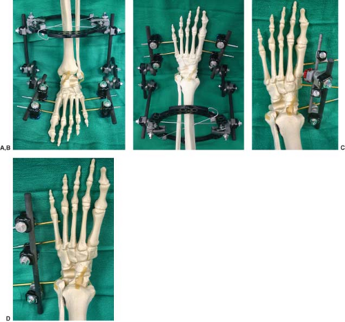 Figure 11.1. The thread diameter of the half-pins to the foot is usually 3 mm for the metatarsals and 4 mm for the midtarsal bones. (A) and (B) show a hybrid configuration of an external fixation system that is used for spanning any midfoot fracture/dislocation. Note the additional tibia external fixation that may consist of one or two circular rings for further stabilization of the lower extremity and its connection to the bar to clamp midfoot apparatus. (C) shows the configuration of a uniplane monolateral external fixation system for spanning an isolated navicular fracture while (D) shows the same principles for spanning a cuboid fracture at the lateral column of the foot. |
Preoperative Considerations
Midfoot Trauma
The initial surgical goal for high-energy midfoot trauma is to maintain the length of medial and/or lateral columns while stabilizing the keystone central column. This can be accomplished with a primary ORIF if the soft tissue envelope permits or with a temporary spanning external fixation to allow for delayed reconstructive procedures if feasible. Pulverized midfoot fractures that result in loss of arch height and/or length of the medial or lateral columns may need to be anatomically reduced while maintaining the functional axis related to the ankle and lower extremity. Articular surface restoration becomes a secondary goal in these pulverized fracture
patterns. Often, these fracture patterns are associated with a compromised soft tissue envelope that requires immediate attention. Stabilizing the foot in its anatomic shape with an external fixator relieves the strain to the surrounding soft tissues and allows for future staged reconstructive procedures to improve further stability and function to the foot (Clinical Case IV).
patterns. Often, these fracture patterns are associated with a compromised soft tissue envelope that requires immediate attention. Stabilizing the foot in its anatomic shape with an external fixator relieves the strain to the surrounding soft tissues and allows for future staged reconstructive procedures to improve further stability and function to the foot (Clinical Case IV).
External fixation may also be utilized as a joint distractor to facilitate ORIF for severe midfoot crush injuries. In these case scenarios, the external fixation is beneficial in achieving fracture reduction and alignment to the columns of the foot. For example, a lateral column spanning external fixator can be utilized as a joint distractor to restore the length in the lateral column of the foot when it is associated with a crushed cuboid.
Alignment of the foot in this manner also facilitates an ORIF of the medial and/or central columns.
Alignment of the foot in this manner also facilitates an ORIF of the medial and/or central columns.
Clinical Case I
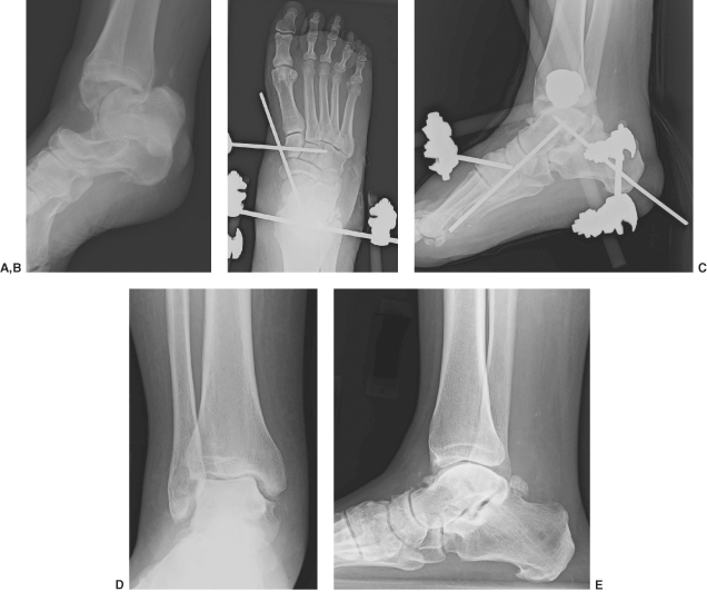
Preoperative lateral radiographic view (A) of a right total talus dislocation. The talus was entrapped between the posterior tibial and flexor digitorum longus tendons preventing a closed reduction. Open reduction and pinning of the peritalar joints was performed followed by the use of an ankle-spanning external fixation for stabilization (B, C). The external fixation system was removed at 6 weeks postoperatively. Final postoperative radiographic views at 8 months showing anatomic alignment of the peritalar joints (D, E). Avascular necrosis of the talus is present without any collapse of the peritalar joints. Serial radiographs are recommended to assess the incidence of talar collapse and posttraumatic arthrosis versus talar revascularization.
 |
Clinical Case II
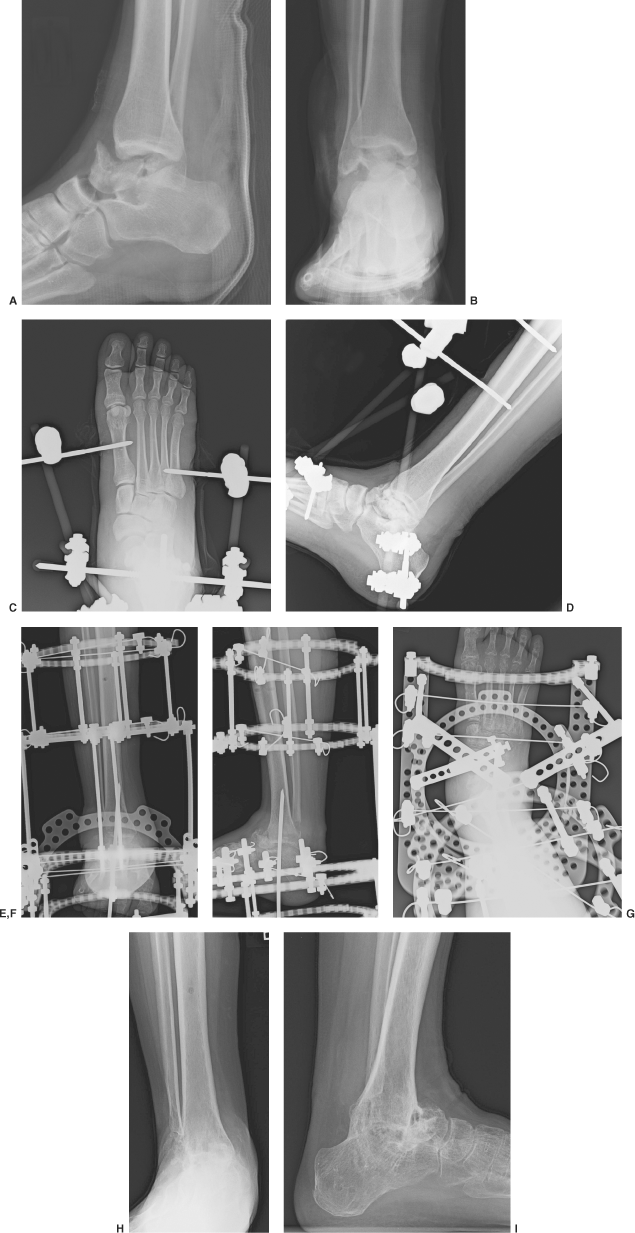
Preoperative lateral (A) and anteriorposterior (B) ankle radiographic views showing a total enucleation to the right talus. The patient originally presented to the emergency room with the talar body retained in a sterile specimen cup and gross contamination from the motor vehicle accident. The patient underwent multiple serial surgical debridements with insertion of cemented antibiotic beads. An ankle-spanning delta external fixator was used to further stabilize the peritalar joints and allow healing of the injured soft tissue envelope (C, D). This temporary external fixation system was removed at 5 weeks postoperatively and was followed by a tibiocalcaneal arthrodesis with a circular external fixation system that consisted of two tibia rings, a foot plate, and an off-loading external ring attached to the foot plate (E–G). The circular external fixator was removed at 14 weeks postoperatively. Final postoperative radiographic views at approximately 6 months demonstrating successful osseous union of the tibiocalcaneal arthrodesis (H, I).
 |
Clinical Case III
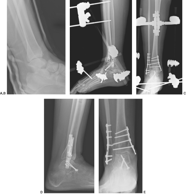
Preoperative lateral radiographic view (A) of a right bimalleolar ankle fracture with an associated talar neck fracture and dislocation at the subtalar joint. An ORIF was performed for the concomitant fractures and an ankle-spanning external fixator was used to further prevent anterior subluxation of the ankle joint by stabilizing the midfoot, hindfoot, and lower extremity (B, C). Final postoperative radiographic views at approximately 4 months demonstrating successful osseous union of the concomitant fractures with anatomic alignment of the peritalar joints and without any evidence of a talar avascular necrosis (D, E).
 |
Clinical Case IV
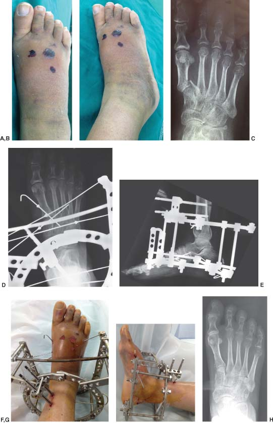
Preoperative clinical (A, B) and radiographic (C) views of a crush injury to the Lisfranc’s joint fixated with the use of a circular external fixator and multiple opposing olive wires. Radiographic (D, E) and clinical (F, G) views at approximately 10 days postoperatively. The external fixation device was removed at 10 weeks with adequate osseous healing and anatomic alignment (H).
 |
Hindfoot Trauma
Partial or total enucleations of the talus commonly present with a transverse to oblique lateral wound associated with a medial talus dislocation. In rare circumstances, the talus can be extruded medially or plantarly through the body of the calcaneus. Lateral talar dislocations are often closed injuries and are not easily amenable to closed reductions as are medial talar dislocations. Often, the talar head lies medial and can be entrapped between the posterior tibial and flexor digitorum longus tendons preventing a closed reduction.
Open wounds associated with a talus dislocation require surgical debridement and irrigation to remove any retained debris that can cause infection. The talus is carefully inspected to determine if any soft tissue attachments remain. For certain case scenarios in which no soft tissue attachments are present within the talus, a decision has to be made in regards to reimplantation and stabilization of the talus to its peritalar joints with external fixation or to perform a talectomy with the insertion of antibiotic-impregnated beads and/or spacer to manage the dead space along with a spanning external fixator. Most commonly reimplantation of the talus is highly preferable unless the open wound is heavily contaminated or the patient is immunocompromised. Close postoperative observation of the reimplanted and fixated talus is paramount in order to reduce any risks of postoperative complications.
External fixation for the management of calcaneal fractures can be quite challenging secondary to the degree of difficulty for joint reduction that can be achieved with the limited placement of half-pins and smooth wires around the fracture pattern. Typically, a half-pin or threaded transcalcaneal pin can be inserted in either the superior or the inferior aspect of the posterior tuberosity to reduce the primary fracture line, medial wall, and/or achieve alignment of the calcaneal tuberosity through traction and manipulation (Clinical Cases V and VI). Another option is to place traction on a half circular ring attached to one or two opposing smooth or olive wires that are placed across the posterior calcaneal tuberosity followed by a circular external fixator to maintain joint reduction (Figure 11.2).
In general, the overall alignment, height, and length of the calcaneal body can be improved with external fixation. In addition, frontal plane alignment of the calcaneal tuberosity can be also reduced with external fixation. In contrast, reduction of the lateral calcaneal wall and/or articular surface of the posterior facet is often only feasible through a limited open reduction for comminuted calcaneal fractures. Tongue-type and two-part fractures involving the posterior facet of the subtalar joint may be reduced with a limited open exposure technique and combined with external fixation (Clinical Case VII).
Other calcaneal fracture patterns often require ORIF to achieve near-anatomic alignment. The technique of external fixation for calcaneal fracture management is not to achieve an anatomic reduction of the articular surface but to reestablish the shape of the calcaneus and to realign the calcaneus to the talus and lower extremity. This technique is utilized to improve the positioning of the calcaneus and allow for osseous consolidation. Improved overall alignment of the calcaneus facilitates future reconstructive surgical options to relieve pain and improve function if deemed necessary.
Detailed Surgical Technique
Stabilizing the Midfoot
An important concept for midfoot stabilization is to have fixation at both proximal and distal regions to the injured site(s). In most cases, midfoot trauma stabilization with external fixation begins with insertion of a transcalcaneal or half-pin in the
calcaneus. A 5 mm threaded transcalcaneal pin is typically utilized if stabilization of both the medial and lateral columns of the foot is needed. The transcalcaneal pin is driven from medial to lateral direction and within the junction of the superior and inferior portion of the posterior calcaneal tuberosity. An alternative 5 mm threaded half-pin can be driven into the posterior portion of the calcaneal tuberosity in a medial or lateral orientation and according to which column of the foot requires stabilization. For the lateral column of the foot, spanning external fixation may include a second half-pin that can be inserted into the proximal region of the anterior process of the calcaneus if not fractured. Typically, a 4 mm threaded half-pin is utilized and a small 0.5 to 1 cm incision is made and carried down to the level of bone below or between the peroneal tendons. Please note that it is important to place a soft tissue sleeve directly down to the level of calcaneus and thus prevent any injury to the sural nerve and/or peroneal tendons. After appropriate fixation is achieved in the calcaneus, half-pin placement is then carried out at the level of the metatarsals. To further stabilize and span the lateral column of the foot, a 4 mm threaded half-pin is then inserted into the base of the fifth and fourth metatarsals. The insertion of the half-pin begins at the proximal region of the metaphyseal/diaphyseal region of the fifth metatarsal and is aimed approximately 15 degrees superiorly to obtain good purchase at the base of the fourth metatarsal. Utilization of a self-drilling half-pin is advantageous as positioning of the appropriate insertion of the half-pin can be confirmed under C-arm fluoroscopy and then driven into the metatarsal bases. Additional stabilization of the lateral column of the foot can be achieved with the insertion of a 3 or 4 mm threaded half-pin into the diaphysis of the fifth metatarsal shaft. Alternative fixation of the lateral column of the foot is the insertion of a 4 mm threaded half-pin into the cuboid if not fractured and for management of a crush injury involving the anterior process of the calcaneus.
calcaneus. A 5 mm threaded transcalcaneal pin is typically utilized if stabilization of both the medial and lateral columns of the foot is needed. The transcalcaneal pin is driven from medial to lateral direction and within the junction of the superior and inferior portion of the posterior calcaneal tuberosity. An alternative 5 mm threaded half-pin can be driven into the posterior portion of the calcaneal tuberosity in a medial or lateral orientation and according to which column of the foot requires stabilization. For the lateral column of the foot, spanning external fixation may include a second half-pin that can be inserted into the proximal region of the anterior process of the calcaneus if not fractured. Typically, a 4 mm threaded half-pin is utilized and a small 0.5 to 1 cm incision is made and carried down to the level of bone below or between the peroneal tendons. Please note that it is important to place a soft tissue sleeve directly down to the level of calcaneus and thus prevent any injury to the sural nerve and/or peroneal tendons. After appropriate fixation is achieved in the calcaneus, half-pin placement is then carried out at the level of the metatarsals. To further stabilize and span the lateral column of the foot, a 4 mm threaded half-pin is then inserted into the base of the fifth and fourth metatarsals. The insertion of the half-pin begins at the proximal region of the metaphyseal/diaphyseal region of the fifth metatarsal and is aimed approximately 15 degrees superiorly to obtain good purchase at the base of the fourth metatarsal. Utilization of a self-drilling half-pin is advantageous as positioning of the appropriate insertion of the half-pin can be confirmed under C-arm fluoroscopy and then driven into the metatarsal bases. Additional stabilization of the lateral column of the foot can be achieved with the insertion of a 3 or 4 mm threaded half-pin into the diaphysis of the fifth metatarsal shaft. Alternative fixation of the lateral column of the foot is the insertion of a 4 mm threaded half-pin into the cuboid if not fractured and for management of a crush injury involving the anterior process of the calcaneus.
Related posts:
 History and Evolution of External Fixation
 Stepwise Approach to Static Circular External Fixation
 Stepwise Approach to Forefoot Elective and Reconstructive Surgery with External Fixation
 Stepwise Approach to Ankle Elective and Reconstructive Surgery with External Fixation
 Surgical Off-loading External Fixation and Plastic Reconstruction of the Foot and Ankle
 Stepwise Approach to Bone Loss, Transport, and Lengthening with External Fixation
Stay updated, free articles. Join our Telegram channel

Full access? Get Clinical Tree


