Abstract
We report the clinical case of a 54-year-old woman presenting radicular low back pain on the right side of L4 associated to spondylolisthesis on L4-L5, without any notion of trauma or spine surgery. Furthermore this patient is regularly seen for benign rheumatoid polyarthritis complicated by steroid-induced osteoporosis. A preventive treatment was implanted with good results on pain improvement and functional capacities. For pedicle fractures the literature review reports several different etiologies: spontaneous fractures, hereditary fractures or stress-related fractures. There was a discussion on the various treatments available and in this case of spondylolisthesis on pedicle fracture a conservative treatment was implemented similar to the one for isthmic spondylolisthesis. It yielded satisfying results.
Résumé
Nous rapportons le cas clinique d’une femme de 54 ans, ayant consultée pour une lombalgie avec une radiculalgie L4 droite en rapport avec une fracture pédiculaire bilatérale de L4 associée à un spondylolisthésis de L4 sur L5, sans notion de traumatisme ni de chirurgie rachidienne. De plus, elle est suivie pour une polyarthrite rhumatoïde bénigne compliquée d’une ostéoporose cortisonique. Un traitement conservateur a été instauré. L’évolution a été favorable avec une amélioration de la douleur et de la capacité fonctionnelle. La revue de la littérature rapporte des étiologies de fractures pédiculaires « spontanées », d’origine congénitale ou en rapport avec des fractures de fatigue. Différents traitements proposés sont discutés et dans ce cas clinique, un traitement conservateur du spondylolisthésis sur fracture pédiculaire identique à celui du spondylolisthésis sur lyse isthmique a permis un résultat satisfaisant.
1
English version
1.1
Introduction
Spondylolisthesis (SPL) is clearly visible on lumbar spine x-ray images often without any clinical explanation . We differentiate two types of SPL, spondylolysis SPL and degenerative SPL. Pedicle stress fractures as well as defects due to congenital aplasia are quite rare etiologies for spondylolysis SPL . Conservative treatment measures are sufficient for most patients (e.g. rest, rehabilitation, analgesics, zygapophyseal joint injections) . We report the case of a patient with low back pain correlated to spondylolisthesis L4–L5 on a bilateral pedicle stress fracture at L4 without any history of spine surgery or trauma.
1.2
Observation
For the past 4 years this 54-year-old female patient has been seen in consultation for mechanical low back pain on her right side, on the visual analogue pain scale (pain VAS) the patient’s pain was at 40 mm. In her history we find benign rheumatoid polyarthritis progressing for the past 9 years, its clinical and structural evolution are positively treated with Disease Modifying Anti-Rheumatic Drugs (DMARDS) and low dose of corticosteroids (between 5 and 10 mg of prednisone per day). She also has steroid-induced osteoporosis (T-score = −1.8 DS). The patient has been menopaused for 4 years. There is no notion of spine trauma or surgery in her medical history.
The progression of the symptoms revealed: a progressive aggravation of the pain evaluated on the pain VAS at 80 mm, a 50-m restricted walking perimeter (WP) and functional impairments evaluated at 70 mm on a VAS.
At the clinical examination we find a positive Leri’s sign on the right side with tactile hypoesthesia and epicritic sensibility in the right L4 nerve root, no motor deficit or lumbar spine syndrome.
The rest of the examination is eventless. There is no biological inflammatory syndrome.
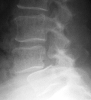
Standard x-ray imaging of the lumbar spine (lateral, anterior-posterior, and oblique views), show a Grade 1 L4-L5 spondylolisthesis according to the Meyerding classification and bilateral pedicle fractures of the L4 vertebra ( Fig. 1 ).

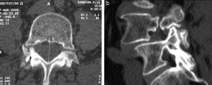
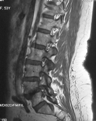
Additional imaging exams (CT-scan and MRI) validate the bilateral pedicle stress fracture, an intact isthmus as well as a right foraminal disc herniation compressing the L4 nerve root on the same side ( Figs. 2 and 3 ).


In the outpatient hospital a conservative treatment was implemented consisting in analgesics belonging to Step II on the World Health Organization pain ladder. A kyphosis-induced rehabilitation program was also prescribed to reinforce the lumbar spine and iliopsoas muscles. A lumbosacral orthosis was ordered with pelvic and sacral support. The patient also benefited from corrective exercises with kyphosis-induced lumbar spine tractions.
The evolution at 1 month reported a 60% global improvement on pain (pain VAS at 30 mm), functional impairment (functional impairment VAS at 35 mm) and walking perimeter (WP = 300 m).
1.3
Discussion
There are several etiologies for spondylolisthesis, we differentiate it into several types: congenital, isthmic, degenerative, traumatic, and pathologic with alteration of the bone structure (e.g. osteoporosis, Paget’s disease, osteolytic tumor) and iatrogenic spondylolisthesis . The lysis of the posterior elements have a significant pathophysiological meaning and unveil, on top of acquired and iatrogenic causes, the congenital defects of the posterior elements .
The pedicle is not the elective location of posterior elements fractures, since they are more common on the isthmus corresponding to the weakest zone . Cyron and Hutton validated this epidemiological observation and reported that posterior elements fractures due to mechanical overuse on the vertebra were located in the isthmus of the pars interarticularis in more than two-third of the cases .
Bilateral pedicle stress fractures of the lumbar spine are quite rare and very few cases were reported in the literature. Most cases are linked to trauma injury, a history of spine surgery or activity-related mechanical stress. Up to then, only four cases of bilateral pedicle stress fractures were described outside of the above-mentioned etiologies. .
Our patient has steroid-induced osteoporosis complicating a rheumatoid polyarthritis making this woman at high risk for stress fractures .
Because of the weak nature of the spinal bones due to osteoporosis, stress fractures can occur after minor trauma or even without any trauma at all .
Sadiq et al. reported the case of a bilateral pedicle stress fractures of the second lumbar vertebra in a 36-year-old patient with a sedentary lifestyle who had chronic lumbar pain of moderate intensity .
Doita et al. reported 2 cases of bilateral pedicle stress fractures. The first case was a 57-year-old male patient with a lumbar canal stenosis with mechanical lumbar back pain and sciatica, imaging of the spine validated a bilateral pedicle stress fracture on L4 . The second case was a 77-year-old female patient with an L5 osteoporotic fracture who complained of intense mechanical lumbar pain on the right side. The CT-scan of the spine validated a bilateral pedicle stress fracture on L4 .
Hyeun Sung Kim et al., reported the case of a bilateral pedicle stress fracture on the second lumbar vertebra with spondylolisthesis in a 63-year-old female patient, treated for ankylosing spondylarthritis with severe chronic lumbar pain .
Our patient did not have any predisposing risk factors for pedicle fractures, the pedicles were intact on a cortical level, so this fracture could be either a stress fracture since the patient has steroid-induced osteoporosis or a fracture due to congenital spine defect. For the latter hypothesis the agenesis seems mainly unilateral .
The four cases of bilateral pedicle stress fractures reported by the literature were treated with surgery . In our case, given that the patient refused the surgery and because it was a low-grade spondylolisthesis a conservative treatment was implemented based on the same therapeutic care used for isthmic spondylolisthesis. To this day, no study has evaluated treatments during the course of pedicle fractures. However several studies evaluated the efficacy of rehabilitation for isthmic spondylolisthesis . Rehabilitation treatment uses various techniques such as kyphosis-induced rehabilitation, reinforcement of the lumbar supporting muscles and sensorimotor reprogramming . Stretching, especially on the lower limbs focuses on treating the retraction of the hamstring muscles, frequently seen in this pathology .
O’Sullivan et al. demonstrated during a prospective, controlled and randomized study that low back pain and functional impairment were significantly decreased in the group that had access to a muscle reinforcement program by co-contraction exercises of the deep abdominal muscles and lumbar spine muscles . We could use pain-relief injections to start the rehabilitation. It is possible to do injections on the posterior joints above the lysis to reach the pedicle “defect” , i.e. infiltrate the L3–L4 joints in case of lysis in L4. Wearing a lombostat orthosis is frequently recommended in athletes but seems poorly indicated in adult patients . Several surgical techniques were proposed to restore lumbar spine anatomy and stability. Arthrodesis followed by the various lumbar spine fusion surgical techniques showed variable biomechanical outcomes and indications according to the patient’s age, SPL’s etiology and impact . However, surgery needs to be considered if the medical treatment is unsatisfactory all the while checking for SPL stability and progression .
Moller and Hedlund did a study on 111 adult patients with isthmic spondylolisthesis with at least a 1-year history of lumbar back pain/sciatica as well as functional impairment. The patients were randomized into two groups: Group 1 patients had posterolateral arthrodesis treatment and Group 2 patients followed a rehabilitation program. Group 1 showed significant improvements on functional impairment and pain compared to Group 2. .
1.4
Conclusion
Bilateral pedicle fractures on spondylolisthesis remain a rare occurrence. Considering all the clinical and imaging data, we agreed on a diagnosis of bilateral pedicle stress fracture. A conservative treatment for these fractures could be discussed first before looking at surgical therapeutic options.
Conflict of interest statement
None.
Acknowledgement
The authors would like to thank Dr. Leila SBIHI for her fruitful partnership.
Stay updated, free articles. Join our Telegram channel

Full access? Get Clinical Tree


