Shoulder


Shoulder


Anatomy


Important areas of shoulder anatomy


  • Musculotendinous units of the rotator cuff and biceps.


  • Bony landmarks of the humerus, scapula, and clavicle.


  • Four joints of the shoulder.


Rotator cuff

The rotator cuff comprises the tendons of four muscles.


  • The subscapularis is located anteriorly on the scapula.


  • The supraspinatus, infraspinatus, and the teres minor are located posteriorly on the scapula.


  • All are closely associated with the glenohumeral capsule, as is the biceps tendon.


  • Primary function of the rotator cuff is to position the humeral head in the glenoid allowing larger muscles to provide necessary power.


Humerus

Important bony landmarks of the shoulder.


  • Greater tuberosity of the humerus:


    • Insertion site of the supraspinatus, infraspinatus, and teres minor tendons.


    • This prominence is often associated with impingement.


    • When testing for impingement on physical exam most tests attempt to force the greater tuberosity under and against the acromion and the coraco-acromial ligament, thus catching or ‘impinging’ the subacromial structures.


  • The bicipital groove is a palpable indentation immediately medial and anterior to the greater tuberosity of the humerus:


    • It houses the tendon of the long head of the biceps.


    • It is easily identified if the humerus is alternately internally and externally rotated, while palpating this area.


    • There is a retinaculum that holds the biceps tendon in place.


  • On the anterior and inferior border of this groove is the lesser tuberosity, the insertion site of the subscapularis.


Articulations

There are three true joints of the shoulder (Fig. 17.2):


  • The glenohumeral, acromioclavicular, and the sternoclavicular joints:


    • The latter two may have a fibrocartilagenous disc present within the articulation.


    • As with all true joints, they are susceptible to various arthritides and trauma.

The fourth joint, the scapulothoracic joint, is a physiological joint, and may be associated with pain syndromes, bursitis, and neuropathies.

The clavicle and its attachments with the sternum, acromion, and coracoid processes are the only bony attachments of the shoulder to the thorax. These connections absorb a large portion of traumatic stresses to the upper extremity and are therefore more susceptible to injuries.


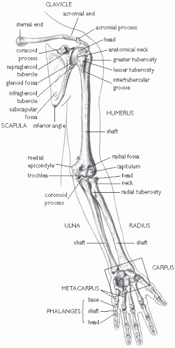
Fig. 17.1 Bones of shoulder girdle and upper limb; anterior view. Reproduced with permission from MacKinnon P and Morris J (2005). Oxford Textbook of Functional Anatomy, Vol. 1. Oxford University Press, Oxford. © 2005.


Glenohumeral joint


  • The glenohumeral joint sacrifices the bony and ligamentous stability of other joints for increased range of motion.


  • The primary stabilizers of the joint are the musculotendinous complex of the rotator cuff and joint capsule, including the labrum.


Bursae

There are 4 important bursae in the shoulder:


  • The subacromial bursa:


    • Located immediately inferior to the acromioclavicular joint and superior to the glenohumeral joint.


    • It is often involved in impingement syndrome.


    • Injection of local anesthetic into this bursa with eradication of symptoms is the basis of the impingement test.


  • The subdeltoid bursa: located inferior to the deltoid tendon on the lateral shaft of the humerus.


  • The subscapular bursa: located between the joint capsule and the tendon of the subscapularis muscle.


  • The subcoracoid bursa: located between the joint capsule and the coracoid process of the scapula.


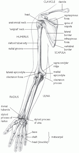
Fig. 17.2 Bones of shoulder girdle and upper limb; posterior view. Reproduced with permission from MacKinnon P and Morris J (2005). Oxford Textbook of Functional Anatomy Vol. 1. Oxford University Press, Oxford. © 2005.


Special tests

The special manoeuvres for evaluation of the shoulder are presented below as described in the literature, and organized according to the problem they evaluate. The reader may have been taught varying or different techniques for the tests presented here. Be cautious in modifying the test, as validity testing will no longer be applicable. A literature review of these manoeuvres is summarized in Table 17.2, but you should be aware that additional well-designed studies are needed in many cases.


Glenohumeral joint stability


Anterior apprehension test

With the scapula stabilized (or in the supine position), the arm is passively moved out to 90° of abduction and gently externally rotated until there is apprehension and the patient resists further ER. The feeling that the joint is going to dislocate (apprehension) constitutes a positive test. Pain alone does not necessarily signify instability. Caution should always be exercised not to completely dislocate the joint with this manoeuvre.


Relocation test

The patient is supine with the arm held in the apprehension position as described above. With a positive test, a posterior force placed on the anterior humerus will relieve the apprehension. Further ER may then be possible. If the posterior force is removed, apprehension returns. This is sometimes called the ‘surprise test’.


Augmentation test (fulcrum test)

To define an occult instability, examiner should place hand underneath the humeral head posteriorly, while the patient is supine and in the apprehension position. This may elicit a positive apprehension test.


Load and shift test

While stabilizing the scapula, the humeral head is loaded medially against the glenoid while in the neutral position. A posterior and anterior stress is applied to the humeral head, as if to shift it anteriorly and posteriorly. Translation of up to 50% of the width of the humeral head is considered normal. Movement to the rim of the glenoid is considered subluxation.


Posterior apprehension

The shoulder is passively moved to 90° of abduction and gently internally rotated until the patient is apprehensive and resists further attempts at IR. A posteriorly directed force placed upon the humeral shaft may intensify the feeling of instability.


Sulcus sign

The arm is passively at the side in the standing or sitting position. The humerus is distracted inferiorly. The sulcus sign is an indentation seen immediately inferior to the acromioclavicular joint and signifies an inferior instability. Normal is less than 1cm. Some patients are able to do this spontaneously as a ‘party trick’. Look for generalized hyperlaxity.


Table 17.2 Shoulder (glenohumeral) joint: movements, principal muscles, and their innervation1


Movement


Principal muscles


Peripheral nerve


Spinal root origin


Flexion


Pectoralis major (clavicular part)


Pectoral nerve (medial and lateral)


C 5, 6


Deltoid (clavicular part)


Axillary nerve


C 5, 6


Extension


Latissimus dorsi


Nerve to latissimus dorsi (thoracodorsal nerve)


C 5, 6, 7, 8


Abduction


Supraspinatus (initial 20°)


Suprascapular nerve


C 5, 6


Deltoid


Axillary nerve


C 5, 6


Adduction


Pectoralis major


Pectoral nerves (medial and lateral)


C 5, 6


Latissimus dorsi


Nerve to latissimus dorsi


C 5, 6, 7, 8


Medial (internal) rotation


Pectoralis major


Pectoral nerves (medial and lateral)


C 5, 6


Latissimuss dorsi


Nerve to latissimuss dorsi


C 5, 6, 7, 8


Subscapularis


Subscapular nerves (upper and lower)


C 5, 6


Teres major


Lower subscapular nerve


C 5, 6


Lateral(external) rotation


Infraspinatus


Suprascapular nerve


C 5, 6


Teres minor


Axillary nerve


C 5, 6


Deltoid (posterior fibers)


Axillary nerve


C 5, 6


Circumduction


Combinations of the above


1Reproduced with permission from MacKinnon P, Morris J (2005). Oxford Textbook of Functional Anatomy Vol 1. Oxford: Oxford University Press, ©2005.


Impingement syndrome


Neer’s sign (impingement sign)

This test attempts to force the greater trochanter under the acromioclavicular joint to compress the bursa, rotator cuff, and biceps tendon. The arm is placed into maximum forward flexion and IR, while stabilizing the scapula. The test is positive if pain is experienced.


Hawkin’s sign

With the elbow flexed at 90° and the shoulder forward flexed at 90° the humerus is progressively internally rotated in order to grind the proximal humerus against the acromioclavicular joint. The test is positive if pain is experienced.


Impingement test

The impingement test involves the injection of 10cm3 of a 1% lidocaine solution into the subacromial space. Greater than 80% resolution of the pain with repeat testing for impingement signs is a positive test suggesting impingement syndrome.


Posterior impingement sign

Executed with the patient supine. The arm is placed in 90-110° of abduction and in slight extension (10-15°). The shoulder is rotated into maximum ER. Recreating symptoms marked by complaints of pain deep within the posterior aspect of the shoulder is indicative of a positive test for posterosuperior glenoid impingement.


Rotator cuff


Drop arm test

With the arm straight, shoulder abducted past 90°, and then forward flexed to 30° the patient is asked to slowly lower it to the side. With a supraspinatus tear he/she is unable to lower the arm slowly and the arm drops to the side.


Jobe’s manoeuvre (empty can test)

With the arm straight, and the shoulder abducted to 90° and horizontally adducted to 30°, the humerus is internally rotated to 45° (as if emptying a beverage can). With a supraspinatus tear, the patient is unable to maintain the position against resistance. The shoulder must be compared with the opposite side. In a recent study, the position of supination (the ‘full’ can position) provided improved isolation of the supraspinatus. It is therefore recommended that both pronation and supination be utilized.


Gerber’s lift off test (lift off test)

With the dorsum of the hand placed over the sacrum, the patient is asked to push away from the back against resistance. With subscapularis weakness or tear, there is weakness on the affected side compared with the contralateral side.


Belly-press test

If the patient is unable to reach the hand behind the back because of restricted range of motion, then it can be placed on the belly instead. The examiner attempts to pull the hand out/away as the patient resists.


External rotator test

With the elbows down at the side and at 90° of flexion, and the humerus in 45° of IR, the patient is asked to externally rotate the humerus against resistance. With infraspinatus weakness there is weakness on the affected side as compared to the contralateral side.


ER lag sign

This tests the integrity of the infraspinatus and supraspinatus. The patient is seated with his/her back to the examiner. The elbow is passively flexed to 90°, with the shoulder held at 20° of abduction in near maximal ER (maximal ER minus 5° to avoid elastic recoil). The patient is then instructed to maintain this position while the examiner releases the arm, supporting only the elbow. The sign is positive if a >10° lag or an angular drop is seen.


The drop sign

This tests mainly the infraspinatus integrity. The patient is seated with his/her back to the examiner. The affected arm is held at 90° of abduction and at almost full ER with the elbow flexed at 90°. The patient is then asked to maintain this position while the examiner releases the hand and supports the arm only at the elbow. The sign is positive if a >10° lag occurs.


The IR lag sign

This tests mainly the subscapularis integrity. The patient is seated with his/her back to the examiner. The affected arm is held behind the back. The elbow is flexed to 90° and the shoulder held at 20° of abduction and 20° of extension. The patient’s hand is then pulled away from the back until it is in almost maximal IR. The patient is then asked to maintain this position as the examiner releases the hand and supports the arm only at the elbow. The sign is positive if a >10° lag occurs.


Acromioclavicular joint


Crossover test (‘scarf’ sign)

With the elbow extended, the arm is brought across the chest stressing the acromioclavicular joint. Tenderness is felt in the superior and lateral shoulder if the test is positive.


Biceps


Yergason’s test

With the elbow at 90° and the wrist held in pronation the patient will experience pain if attempting to supinate the wrist against resistance.


Speed’s test

With the elbow flexed at 30°, the shoulder at 60° of flexion and the wrist supinated, the patient will experience pain if attempting to flex the arm against resistance.


Labral tears


Biceps load test II

With the patient in the supine position the examiner holds the patient’s wrist and elbow with the shoulder in 120° of abduction and maximally externally rotated. The elbow is placed in 90° of flexion and the forearm supinated. The patient is asked to flex the elbow against the examiner’s resistance. The test is considered positive for a labral lesion if there are increased complaints of pain with resisted elbow flexion.


Anterior slide test (Kibler)

The patient is examined either in a standing or sitting position, hands on the hips and thumbs pointing posteriorly. One of the examiner’s hands is placed across the top of the shoulder from the posterior direction, with the index finger over the anterior acromion. With the other hand, the examiner applies a forward and slightly superiorly directed force to the elbow and upper arm. The patient is asked to push back against this force. Pain localized to the front of the shoulder under the examiner’s hand and/or a pop or click in the same area, is considered a positive test.


Clunk test

While applying gentle axial pressure to the humerus with the elbow at 90°, the humerus is rotated internally and externally while simultaneously abducting the arm. If the examiner’s opposite hand is placed underneath the humeral head, a clunk, pop, or snap may be felt if a labral tear is present.


Crank test

With the patient in the sitting or standing position, the arm is elevated to 160° in the scapular plane. An axial load is applied to the humerus, while simultaneously internally and externally rotating the humerus in the glenoid fossa. Pain or reproduction of the patient’s symptoms (usually pain or catching) is considered a positive test.


O’Brien test

Performed with the patient in the sitting position, the shoulder is abducted to 90° with the elbow in full extension. The forearm is then pronated with the thumb pointing down. A downward force is placed on the arm by the examiner. The force is repeated with the forearm in the supinated position with the thumb pointing upwards. Pain or clicking inside the shoulder joint is considered a positive test for a labral lesion. Pain more superficially, over the acromioclavicular joint indicates pathology in that joint.


Thoracic outlet syndrome


Adson’s manoeuvre

While feeling the radial pulse the arm is passively abducted, extended, and externally rotated (simultaneous abduction and ER of the arm compresses the brachial plexus against the scalene muscles). The head is extended and turned to the side of the lesion, while the patient holds his/her breath. Loss of or a diminished radial pulse is a positive test.


Wright’s manoeuvre (hyperabduction manoeuvre)

Similar to Adson’s manoeuvre, but the arm is hyperabducted over the head, while externally rotated and extended (this simulates compression of the neurovascular bundle beneath the pectoralis tendon). Loss of or a diminished radial pulse is a positive test.


Costoclavicular manoeuvre (military brace position)

The patient sits upright and thrusts the shoulders backward, while the hands rest on the thighs. This narrows the space between the clavicle and the first rib. This may reproduce the symptoms if a ‘backpacker’s neuropathy’ is present.


Roos test (overhead exercise test)

The arms are abducted to 90°, shoulders externally rotated, and the elbows flexed to 90°. The hands are opened and closed for 3min to reproduce the symptoms. This may be positive in baseball pitchers with symptoms while in the cocking phase (though this mechanism may also represent an anterior instability). In extreme cases, there may also be blanching of the affected hand(s) with this test.


Subscapular bursa


  • Located between the scapula and the thoracic wall.


  • May be multiple small bursae.


  • May become inflamed with over-use, causing ‘snapping scapula’.


  • Often difficult to diagnose.


  • May need to managed surgically if persistent symptoms.


Nerves

The brachial plexus is a complex array of roots, trunks, divisions, cords, and branches.


  • Any of these areas may be injured with compression or stretching of the brachial plexus.


  • Because of the high incidence of anterior shoulder dislocations, the most likely peripheral nerve to be damaged is the axillary nerve.


  • Other peripheral nerves that are likely to be damaged include the suprascapular, musculocutaneous, and the long thoracic nerves.


Shoulder disorders


Epidemiology


  • Shoulder pain is the second most common musculoskeletal complaint seen by practitioners: 1.2-2.5% of attendances in primary care.


  • Rotator cuff lesions (65%).


  • Pericapsular soft tissue pain (11%).


  • Acromioclavicular joint pain (10%).


Factors related to early recovery


  • Mild trauma.


  • Acute onset.


  • Over-use problems.


  • Early presentation.


Problems that are related to prolonged recovery


  • Diabetes mellitus.


  • Cervical spondylolysis.


  • Radicular symptoms.


  • Advancing age.


  • Involvement of the dominant extremity.


Causes

Causes of increased susceptibility to injury include:


  • An inherently less stable joint.


  • The fact that we ‘abuse’ the joint with repetitive work activities and participate in sports that push the limits of the joint.


Differential diagnosis

Acute problems with rapid onset over days to a few weeks include:


  • Trauma.


  • Acute over-use.


  • Cervical nerve root compression.

Age of the patient and history may narrow the differential diagnosis:


  • Patients below the age of 45 often have a biomechanical cause to their problem, such as instability or tendinopathy..


  • Those older than 45 are more likely to have degenerative conditions, such as osteoarthritis or rotator cuff tears.


  • Evaluate the patient for adhesive capsulitis if there is a history of diabetes mellitus, progressive pain, and loss of motion.


Acute traumatic causes


Anterior glenohumeral instability


  • From grade I, with less than 50% subluxation of the humeral head beyond the glenoid fossa, to grade IV, which is a complete dislocation.


Anterior dislocation


  • If a patient lands on the posterior aspect of the shoulder during a fall forcing the humeral head anteriorly on the glenoid or if there is forced ER, while the upper extremity is abducted.


  • The pressure of the anterior glenoid on the posterior humerus during dislocation may cause a small compression fracture or divot on the posterior humeral head known as a Hill—Sach’s lesion.


  • While the humeral head is being forced anteriorly, the anterior capsule may tear the labral cartilage away from the underlying glenoid, termed a Bankart lesion, worsening the anterior instability.


History and examination


  • Patients usually present with pain in the anterior and lateral shoulder.


  • They will often complain of an inability to move the shoulder without significant pain and, in many cases, ‘know’ it is dislocated.


  • With an acute anterior dislocation, the acromioclavicular joint may be prominent on exam, and the shoulder appears ‘sunken’.


  • Patients will hold the arm in a partially abducted and externally rotated position.


  • If the shoulder has relocated, they may have signs of instability on examination, or a positive Speed’s test or apprehension test.


Investigation


  • Diagnosis of dislocation is usually obvious on inspection.


  • May be identified by X-ray evaluation, particularly the scapular Y view.


  • A thorough neurovascular exam should be documented in all cases.


Posterior glenohumeral dislocation

Posterior dislocations account for only 4% of dislocations. Posterior dislocations generally require great force and are often seen after motor vehicle accidents, seizure activity, repetitive weight lifting, or in football linemen. The mechanism is usually a force applied to the forward flexed upper extremity at 90°, forcing the humerus posteriorly. It may also occur
after an anterior blow to the shoulder. A small avulsion of the posterior glenoid labrum may occur resulting in a reverse Bankart lesion.


History and examination

Jul 22, 2016 | Posted by in ORTHOPEDIC | Comments Off on Shoulder

Full access? Get Clinical Tree

Get Clinical Tree app for offline access