Rheumatoid Arthritis
Joan M. Von Feldt
Rheumatoid arthritis (RA) is the most common autoimmune rheumatic disease, affecting 1% to 2% of the population.1 It affects women more than men in a ratio of 5:1 with a peak age of onset between 30 and 55. Untreated, it can cause deformity, significant disability, and premature death.2,3 Although there is no cure for RA, there has been an explosion of new treatments and combination therapies available to treat RA, and many patients can achieve near remission with aggressive therapeutic regimens.
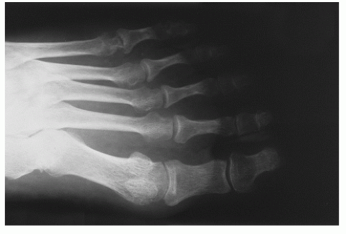
The characteristic findings in RA include inflammatory arthritis that is destructive to joints. This destruction is measured as erosions and joint space narrowing. The destructive arthritis is also accompanied by muscle atrophy, and inflammation of the tendon sheaths that can lead to tendon rupture. Structural damage to the joints and supporting ligaments and tendons is usually irreversible. The degree of damage is closely linked to inflammation and hence to disease activity, but is also associated with degeneration and repair. Extraarticular manifestations can be present as well.4 RA, like other systemic inflammatory conditions, is associated with premature mortality, usually attributed to cardiovascular events.3,5 Therefore, early recognition of the disease and aggressive intervention are warranted.
CLINICAL PRESENTATION
The typical presentation is a young woman with morning stiffness of multiple joints of the hands, wrists, and feet, although almost any joint can be involved in RA. Morning stiffness is usually more prominent than pain. Frequently, women present with chronic foot pain, which they incorrectly attribute to poor shoe wear. Subcutaneous nodules (rheumatoid nodules) can be present on extensor surfaces, although they can be found in lung and other tissues as well. On exam the involved joints are usually swollen, painful on range of motion, and tender, but the findings are usually more subtle than, for example, an inflamed joint affected by gout (Figs. 62-1 and 62-2).
There are new 2010 ACR-EULAR classification criteria for the diagnosis of RA (Table 62-1).6,7 Typically in rheumatic diseases, criteria are labeled as “classification” criteria, not diagnostic criteria. A clinical diagnosis has to be established by the physician (usually a rheumatologist) and may include many more aspects than can be included in formal criteria. With the new criteria, the definition of “small joint” includes the metacarpophalangeal (MCP), proximal interphalangeal (PIP), metatarsophalangeal (MTP) 2 to 5, thumb interphalangeal, and wrist but not distal interphalangeal (DIP), first carpometacarpal, and first MTP. The definition of “large joint” includes shoulder, elbow, hip, knee, and ankles. The criteria for the definition of serology include negative: ≤ULN (for the respective lab); low positive: >ULN but ≤3 ×ULN; and high positive: >3 × ULN. Although erosions and joint space narrowing are a hallmark of the disease, radiographs are not required in the ACR/EULAR 2010 classification criteria.
More frequent extra-articular features of RA include anemia, serositis (pleuropericarditis), scleritis, and secondary Sjögren syndrome (dry eyes and dry mouth). Less commonly, splenomegaly, vasculitis, neuropathy, interstitial lung disease, and renal disease may occur during the course of the disease and can be life threatening. Neck pain should raise concern because of possible involvement of the atlantoaxial joint of the cervical spine and subsequent neck instability. Prompt imaging should be considered.
CLINICAL POINTS
The condition is the most common autoimmune rheumatic disease.
Women are affected more often than men.
Inflammatory arthritis that destroys joints is characteristic.
STUDIES (LABS, X-RAYS)


