Replacement Arthroplasty in Synovial-Based Arthritis
Replacement Arthroplasty in Synovial-Based Arthritis
I. A. Trail
INTRODUCTION
Despite many recent advances in the medical treatment of inflammatory arthritides there are still large numbers of patients with these conditions who present for surgical treatment. The definition of “synovial-based” arthritis is in itself controversial. However, for most clinicians this would mean both seropositive and seronegative rheumatoid arthritis; the latter would include conditions such as gout, chondrocalcinosis, ankylosing spondylitis, and psoriasis. This list is by no means exhaustive, however, and others will be added as time progresses. In practical terms, particularly with regard to replacement arthroplasty, this group would include all patients who do not suffer from osteoarthritis (either primary or secondary), avascular necrosis, or the consequence of trauma. The principal difference is that in synovial-based arthritis the conditions tend to affect a younger age group, occur predominately in females, and usually result in a poorer outcome. On review of literature it is difficult to identify when the first shoulder joint replacement was undertaken for inflammatory arthritis; undoubtedly it has formed the mainstay of treatment for this condition for many years.
The aim of this chapter is to identify the particular and peculiar characteristics of this condition together with the role of arthroplasty. Particular emphasis will be placed on the differences with regard to joint replacement in osteoarthritis.
SURGICAL ANATOMY
The anatomy of the shoulder in patients with synovial-based arthritis can vary immensely. In some circumstances it can be very similar to osteoarthritis; in others, and more commonly, extensive soft-tissue and bone erosion can be seen. This is very much dependent on the stage of the disease process at the time of surgery. With regard to rheumatoid arthritis the condition is known at least initially to be predominately a synovitis that results in capsular distension, soft-tissue damage, and ultimately instability—the latter occurring as a direct result of the ligamentous attenuation and posture. The two most important soft-tissue structures at risk are first, the rotator cuff and second, the long head of biceps.
With regard to the rotator cuff it should be noted that in almost all cases there is some involvement. This involvement is quite different from the degenerative changes seen in rotator cuff tendonopathy/impingement. Most specifically there is a widespread thinning or attenuation of the cuff, and only rarely do you see small- or medium-sized identifiable tears. At the end stage of the disease, however, massive tears are frequently seen. Of further note is the so-called “nonfunctional” cuff that is seen in this type of inflammatory condition (i.e., although a thinned attenuated structure is seen at surgery clinically, the patient does not demonstrate any cuff function). For this reason it is felt that the cuff ceases to function before finally rupturing. The exact pathophysiology of this and its timing in the disease process remains both difficult to determine and controversial.
With regard to the biceps tendon, again this is frequently involved in the synovitic process, resulting in weakness and ultimately rupture, usually at the insertion of the long head into the superior labrum (Fig. 9-1). The effect of this on shoulder function remains controversial; certainly it does not appear to have any major impact on elbow flexion. With regard to the glenohumeral joint, opinion varies as to the importance of the long head of biceps (i.e., it acts as a depressor and stabilizer of the humeral head or it is of little bearing). Certainly there are surgeons who routinely perform a biceps tenotomy in the presence of any pathology. What can be said with more certainty, however, is that rupture of the long head of biceps in patients with synovial-based arthritis appears to have little clinical effect, whereas involvement of the rotator cuff almost certainly does.
Figure 9-1 Arthroscopic view of biceps rupture.
Ultimately the synovial process begins to involve bone and the articular surface. Initially this attacks the rim of the humeral head and glenoid, resulting in periarticular erosions. Further to this, there is generalized humeral head and glenoid articular cartilage damage. This takes the form of fibrillation and ultimately cartilage loss and bone erosion. The pattern of cartilage loss and bone erosion can be quite variable; although commonly central, anterior, and superior glenoid cartilage and bone loss are seen. The anterior-superior loss appears progressive, being noted by Lehtinen and colleagues (1) in their long-term follow-up study of patients affected by rheumatoid arthritis of the shoulder and associated with involvement of the rotator cuff. Over 15 years they noted most severe involvement of the glenohumeral joint in patients who had a narrowing of the acromio-humeral distance, the latter indicating severe rotator cuff involvement.
For the glenoid, although again patterns of erosion can vary, the most frequently seen superior and anterior patterns directly mirror wear in the humeral head. As part of this the humeral head can ultimately sublux anteriorly as well as superiorly. In extreme cases the humeral head can be palpated directly under the skin anterior to the acromion.
PATHOPHYSIOLOGY
The exact incidence of shoulder involvement in patients with rheumatoid arthritis remains unknown. A review of all standard textbooks reveals figures ranging from 20% to 50%. However, most of these figures are based on studies of patients at one point of time in the disease process. Long-term prospective studies would indicate a higher percentage, although obviously the majority of these patients do not require surgical intervention.
The typical clinical picture is one of a patient complaining of pain and stiffness of the shoulder. Although this may follow a minor traumatic event in the majority of cases, there is often no obvious participating cause. It is not unknown in my experience for the disease to present primarily in the shoulder region. Indeed, it is well known that spontaneous pain and swelling in a sternoclavicular joint often heralds the onset of a generalized inflammatory condition. Once the diagnosis is made and medical treatment is begun, the condition of the shoulder can fluctuate; whether the shoulder becomes more seriously involved appears to be the result of nothing more than pure chance. That is not to say that the condition cannot be influenced by activity. Patients who undertake prolonged and continued activities with the arms elevated or who spend long periods on crutches will undoubtedly suffer more with their shoulders. Ultimately, however, if the condition does progress, pain will continue and movements will become markedly restricted. The shoulders themselves will become quite stiff. This combination results in significant loss of function, not only in the shoulder but the upper limb in general.
TABLE 9-1 RADIOGRAPHIC EVALUATION OF RHEUMATOID ARTHRITIS AND RELATED CONDITIONS BY STANDARD REFERENCE FILMS
Grade 0
Normal conditions. Abnormalities not related to arthritis, such as marginal bone deposition, may be present.
Grade 1
Slight abnormality. One or more of the following lesions are present: periarticular soft-tissue swelling, periarticular osteoporosis, and slight joint space narrowing.
Grade 2
Definite early abnormality. Erosion and joint space narrowing corresponding to the standards. Erosion obligatory except in the weightbearing joints.
Grade 3
Medium destructive abnormality. Erosion and joint space narrowing corresponding to the standards. Erosion is obligatory in all joints.
Grade 4
Severe destructive abnormality. Erosion and joint space narrowing corresponding to the standards. Bone deformation is present in the weightbearing joints.
Grade 5
Mutilating abnormality. The original articular surfaces have disappeared. Gross bone deformation is present in the weightbearing joints. Dislocation and bony ankylosis, being late and secondary, should not be considered in the grading; if present, the grading should be made according to the concomitant bone destruction or deformation.
Larsen A, Dale A, Eek M. Radiographic evaluation of rheumatoid arthritis and related conditions by standard reference films. Acta Radiologica Diag 1977;18:481.
CLASSIFICATION
A comprehensive review of the classification of rheumatoid arthritis remains outside the remit of this chapter. Rheumatologists have devised various criteria to help them with diagnosis. These have been expanded to cover seropositive and seronegative varieties depending on the presence of positive serology and the degree of involvement, specifically unilateral or diffuse joint involvement. Seronegative inflammatory arthritis is further subdivided into erosive and nonerosive types. Finally, there are various other terms used such as rheumatoid nodulosis and palindromic rheumatism. Most surgical classifications for rheumatoid arthritis of the shoulder, however, are based on x-ray appearances; the most widely used was described by Larsen and others in 1973 (Table 9-1), which is common to all joints. This method of assessment has been validated (2).
Crossan and Vallance, in 1982, reported their analysis of 100 shoulder joints in patients with rheumatoid arthritis and came up with the grading system shown in Table 9-2 (3). They also correlated the x-ray appearances with the clinical features and found deterioration in pain, movement, and function with increasing grade. They also looked at the significance of the subacromial distance and found a significant reduction below 6 mm between Grades 2 and 3. Finally they also looked at progression and found this occurred at least radiologically in 75% of cases over the study period. They typically reported both superior and medial migration of the humeral head—the former presumably as a result of rotator cuff wear or rupture and the latter as a result of bony erosion of the glenoid and humeral head.
Lehtinen and colleagues (4), in their 15-year follow-up of 148 rheumatoid shoulders, found that medialization actually proceeded upward migration of the humeral head, indicating bone erosion proceeded rotator cuff damage. In light of this they recommended an orthopedic opinion at an early stage.
One other traditional method of classification relies on the phase or stage of the inflammatory disease (i.e., whether there is a synovial reaction or whether the disease is “burned out”). The former is often known as the “wet” stage and the latter is known as the “dry” stage. The surgical relevance is that the former is often treated by synovectomy or other soft-tissue procedures, whereas the latter can be treated in a similar fashion to degenerative arthritis. Levigne and Franceschi (5) quoted incidences of wet and dry stages of 19% and 36%, respectively. They also added a “resorptive” group with superior migration of the humeral head and erosion of the superior aspect of the glenoid; this accounted for 41%.
EVALUATION
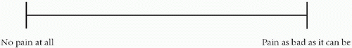
The clinical evaluation of any painful joint falls into three areas. These are pain, function, and all “objective” clinical signs. Pain remains the most important and is the main indication for intervention. It is usually measured by a visual analogue scale in various formats. Generally a simple 0 to 10 assessment of pain is sufficient, however. Function can be assessed in various ways by a series of questions that assess an ability to undertake activities that require either movement or strength. Objective signs include measuring range of motion and strength. These measurements can then be amalgamated into a score; the most common two used are the American Shoulder and Elbow Surgeons (ASES) (Table 9-3) and, in Europe, the Constant-Murley (Table 9-4) scores (6). Although these undoubtedly simplify evaluation and its representation, they do not always reflect the true disability and the outcome of various treatment modalities for specific conditions (e.g., instability).
TABLE 9-2 CROSSAN AND VALLANCE RADIOLOGIC ASSESSMENT OF RHEUMATOID ARTHRITIS
Grade 1
Normal
Grade 2
Periarticular erosions
Grade 3
Proximal subluxation of humeral head, erosive changes
Grade 4
Loss of glenohumeral space
Grade 5
Partial destruction of humeral head and glenoid fossa, loss of sphericity
In addition they poorly reflect the differences in expectation between various patient groups (e.g., sports man versus disabled person). Patients with an inflammatory synovitis affecting several joints in the upper limb will obviously not have as good an outcome following arthroplasty as a patient with single joint disease. In addition, recent research has indicated that, at least in cases of inflammatory arthritis, the scores produced by the ASES score and the Constant-Murley score are not directly interchangeable in so much as the ASES produces a higher value in the same patient as the Constant-Murley. Given these difficulties, many researchers favor the Disabilities of the Arm, Shoulder, and Hand (DASH) score (Table 9-5) (7), which reflects disability of the whole limb in this disease group.
Finally outcome is also affected by the general health of the patient; for this reason many authors believe the short form 36 (SF36) is sufficient. Its particular advantage is that it can be administered as a postal questionnaire. At this time much work is being undertaken in this area, specifically by statisticians, and hopefully there will be some consensus in due course.
Radiologic evaluation has been based on plain x-rays undertaken with the arm in various positions. Indeed, as has been described, many if not all of the classifications have been based on this modality. Computed tomography undoubtedly gives better images of the bone and can be an invaluable adjunct to surgery, particularly in planning glenoid surgery (Fig. 9-2). Magnetic resonance is now being used more frequently and allows better visualization of the soft tissues. This will aid more and more with the planning and execution of the nonarthroplasty procedures.
TABLE 9-3 AMERICAN SHOULDER AND ELBOW SURGEONS SHOULDER SCORE
In the following activities of daily living, please describe how well you can use your OPERATIVE SHOULDER.
Please describe your OPERATIVE SHOULDER on the following scale by placing a vertical line through the part of the scale that you feel is most appropriate.
How bad is your shoulder pain today?
TABLE 9-4 CONSTANT MURLEY SHOULDER SCORE
The Constant Shoulder Assessment Scoring for the following:
1.
Pain
Max = 15
Points
None
15
Mild
10
Moderate
5
Severe
0
2.
Activities of Daily Living Max = 20
Points
Activity level:
Full work
4
Full recreation/sport
4
Unaffected sleep
2
Positioning:
Up to waist
2
Up to xiphoid
4
Up to neck
6
Up to top of head
8
Above head
10
3.
Forward and Lateral Elevation
Elevation (degrees)
Points
0-30
0
31-60
2
61-90
4
91-120
6
121-150
8
151-180
10
4.
External Rotation Max = 10
Points
Hand Position
2
Behind head, elbow forward
2
Behind head, elbow back
2
Top of head, elbow forward
2
Top of head, elbow back
2
Full elevation from top of head
2
5.
Internal Rotation Max = 10
Dorsum Hand Position
Points
Lateral High
0
Buttock
2
Lumbosacral junction
4
Waist (L3 vert)
6
T12 vert
8
Interscapular
10
6.
Overall Scoring for Individual Parameters
Max
Right
Left
Pain
15
Activities of daily living
20
Range of motion
40
Power
25
Total
100
SURGICAL MANAGEMENT
Indications for Surgery and Alternatives to Arthroplasty
The principal indication for surgical intervention is the failure of medical treatment (i.e., despite appropriate medical treatment in the form of drug therapy, intraarticular injection, and other modalities the patient continues to experience significant pain, stiffness, and loss of function). The principal indication for surgery, however, is always pain, particularly if it requires considerable analgesia to control, results in sleep disturbance, and significantly affects a patient’s quality of life.
With regard to the various procedures available it is important to remember that arthroplasty is not the only procedure undertaken in that a number of authors have shown the advantages of synovectomy, either open or by arthroscopic means, in the earlier stages of the disease as well as procedures directed to the subacromial space, rotator cuff, and acromioclavicular joint. Initially Smith-Peterson (8), then Pahle and Kvarnes in 1985 (9), reported the results of open synovectomy in patients with rheumatoid arthritis after a mean follow-up of 5.3 years. Originally they performed a more extensive technique that involved resection of the acromion, after release of the deltoid muscle, and splitting the rotator cuff. More recently, however, the technique has changed in that the resection of the acromion is no longer undertaken. They identified better results in the earlier stages of the disease process; only 6 patients out of 54 went on to total joint replacement.
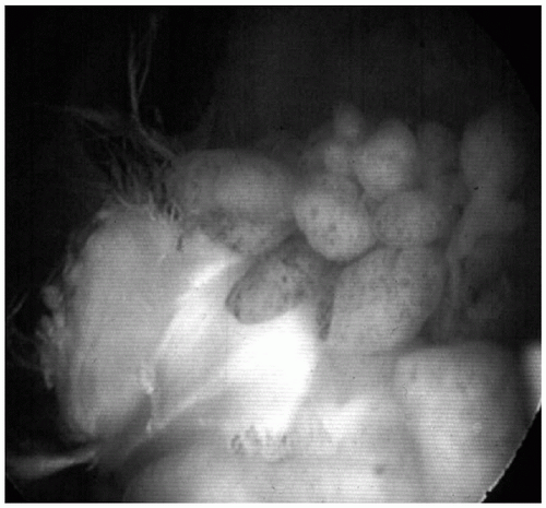
With regard to arthroscopic synovectomy at my institution, this procedure has been undertaken for several years, generally at the behest of rheumatologists who believe they can no longer control the patient’s symptoms; the patients have near normal x-rays. The technique developed uses a fluid management system, shavers, and so on, and has proved just as easy, if not more so, than the conventional open techniques (Fig. 9-3). Results to date have shown good response with regard to pain relief, although no major long-term improvements in range of motion have been observed. Out of 22 patients, only one has gone on to total joint replacement.
Only gold members can continue reading. Log In or Register to continue