Replacement Arthroplasty in Glenohumeral Arthritis: Intact or Reparable Rotator Cuff


Replacement Arthroplasty in Glenohumeral Arthritis: Intact or Reparable Rotator Cuff


Crystal A. Smith

Gerald R. Williams Jr.


INTRODUCTION

Prosthetic replacement for glenohumeral arthritis was first performed by Pean in 1893 in a patient with tuberculosis of the shoulder (1). The device was constrained and failed 2 years after implantation because of recurrent infection. However, the modern era of shoulder replacement with unconstrained, anatomically designed implants began in the mid to late 1950s (2, 3, 4). The first reported results following shoulder replacement for glenohumeral arthritis in
a large series of patients was by Neer in 1974 (5). All patients had glenohumeral osteoarthritis with an intact or reparable rotator cuff. All but one of the patients underwent humeral hemiarthroplasty, with the remaining patient receiving a cemented all-polyethylene glenoid component. Of 48 shoulders, 42 attained satisfactory results.

Neer’s initial success with unconstrained shoulder arthroplasty has continued (6). However, the functional results of shoulder replacement are variable according to the disease process for which the replacement is performed. Conditions characterized by an intact or reparable cuff provide the best functional results. Conversely, conditions with severe or irreparable rotator cuff deficiency, such as cuff-tear arthropathy or rheumatoid arthritis, yield less satisfactory functional results. Primary glenohumeral osteoarthritis and nontraumatic avascular necrosis (AVN) are the prototypical conditions characterized by an intact or reparable rotator cuff and most often produce the best results following prosthetic replacement. Moreover, the operative indications and surgical principles are similar in these two conditions. Therefore, they are considered together in this chapter.

As in other joints and in other disease processes, arthroplasty becomes an option when all attempts at conservative treatment have failed. Nonsurgical management and surgical alternatives short of prosthetic replacement should be considered the mainstay of treatment in patients with mild or moderate degenerative change, whether it is from primary osteoarthritis or AVN. Generally accepted indications for shoulder replacement include severe pain that is unresponsive to conservative management and/or an unacceptable degree of functional impairment. Patients who have had previous surgery, have a history of local infection, or who are younger than 50 years old require careful consideration prior to undertaking this type of treatment. In addition to specific surgical indications, this chapter discusses the surgical anatomy, pathophysiology, diagnostic evaluation, surgical approaches, and results of prosthetic replacement for primary osteoarthritis and nontraumatic AVN of the shoulder.


SURGICAL ANATOMY


Normal Anatomy

Although glenohumeral anatomy is described in detail in Chapter 2 of this text, certain points relevant to replacement for osteoarthritis and AVN will be emphasized here.

The central 80% of the humeral head is spherical, and the peripheral 20% is elliptical (7). However, if one assumes that the entire articular surface is spherical, humeral head radius of curvature, humeral head thickness, retrotorsion with respect to the humeral shaft, and neck-shaft angle are extremely variable (7, 8, 9, 10). Mean humeral head radius is approximately 24 mm, with a range of 19 to 28 mm (7,10,11). Mean humeral head thickness is approximately 19 mm, with a range of 15 to 24 mm (7,10,11). Both humeral head radius and thickness correlate strongly with humeral shaft length and patient height (7). However, the ratio of humeral head thickness to humeral head radius of curvature is remarkably constant at approximately 0.7 to 0.9, regardless of patient height or humeral shaft size (7,10,11). The surface arc of the humerus available for contact with the glenoid is directly proportional to the ratio of humeral head thickness to humeral head radius and is, therefore, also relatively constant (11).

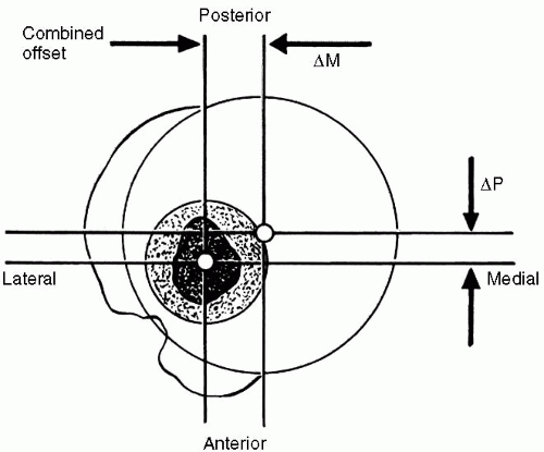
The distance between the center of the humeral head and the central axis of the intramedullary canal is defined as the humeral head offset (8,11,12). Although humeral head offset is undoubtedly three-dimensional, it is commonly described in two planes, coronal and axial. Similar to most other proximal humeral anatomic parameters, reported humeral head offsets are variable (8,11,12). In the coronal plane, the humeral head offset is approximately 7 to 9 mm medial to the central axis of the intramedullary canal; in the axial plane, the humeral head offset is 2 to 4 mm posterior to the central axis of the intramedullary canal (Fig. 7-1) (8,11,12).

Humeral head offset is correlated with humeral head radius and humeral head thickness. However, for a given humeral head radius, humeral head thickness, and humeral head offset in the coronal and axial planes, the location of the humeral articular surface may vary relative to angle of rotation about the central intramedullary axis (i.e., humeral retrotorsion). Humeral retrotorsion averages 20 to 30 degrees, with a wide range of approximately 20 to
55 degrees (8,10, 11, 12). The vertical distance between the highest point of the humeral articular surface and the highest point of the greater tuberosity (i.e., head to greater tuberosity height) is approximately 8 mm and shows a relatively small range of interspecimen variability (7,11).


Figure 7-1 The humeral head center is offset 2 to 4 mm posterior and 7 to 9 mm medial to the central axis of the humeral shaft. (From Boileau P, Walch G. The three-dimensional geometry of the proximal humerus. Implications for surgical technique and prosthetic design. J Bone and J Joint Surg-Br 1997;79(5):857-865, with permission.)

The neck-shaft angle is defined as the angle subtended by the central intramedullary axis of the humeral shaft and the base of the articular segment. The average neck-shaft angle is 40 to 45 degrees (7,11,12). However, more importantly, humeral neck-shaft angle demonstrates significant individual variation with a range of 30 to 55 degrees (7,11,12).

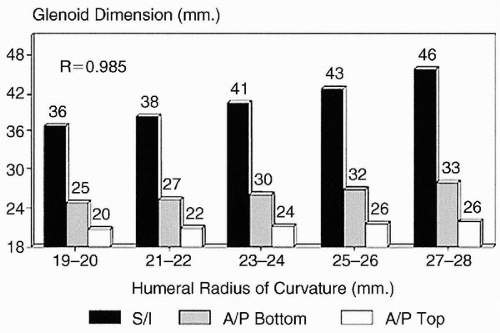
The size and shape of the articular surface of the glenoid can be defined by its linear superoinferior and anteroposterior dimensions, as well as by its radius of curvature (7,13). The mean superoinferior dimension of the glenoid (excluding the labrum) is approximately 39 mm (range 30 to 48 mm) (7,13). The anteroposterior dimension of the superior half of the glenoid is shorter than the inferior half of the glenoid, resulting in a pear-shaped appearance. The mean anteroposterior dimension of the superior half of the glenoid (excluding the labrum) is approximately 23 mm (range 18 to 30 mm), and the mean anteroposterior dimension of the inferior half of the glenoid (excluding the labrum) is approximately 29 mm (range 21 to 35 mm) (7,13). The ratio of the superoinferior dimension to the anteroposterior dimension of the larger, inferior half of an average glenoid is 1:0.7 (7). The humeral head radius correlates with the size of the glenoid in both the superoinferior and anteroposterior dimensions (Fig. 7-2) (7).

Controversy exists about the relationship between glenoid and humeral articular radius of curvature (7,14, 15, 16). This controversy exists because of differences in measuring techniques, differences in sample sizes, and large individual variations in anatomy. The thickness of the articular cartilage of the glenoid increases toward the periphery of the articular surface and must be included when measuring the glenoid radius of curvature (16). However, even when the articular cartilage is included in the measurement, the radius of curvature of the glenoid articular surface does not equal the radius of the humeral articular surface in all specimens (7,14, 15, 16). Iannotti and colleagues (7) observed that, on average, the glenoid radius of curvature in the coronal plane was 2.3-mm larger than the coronal plane humeral radius of curvature in the same specimen. Soslowsky and coworkers (16) reported a difference between humerus and glenoid radii of curvature of less than 2 mm in 88% of specimens and less than 3 mm in all specimens. Kelkar and associates (14,15) reported a mean humeral radius of curvature that was 2 mm less than the mean glenoid radius of curvature.

Normal glenoid version also exhibits significant individual variation (17, 18, 19). Furthermore, the amount of measured retroversion can vary depending on the method of measurement and with the portion of the glenoid being measured. If computed tomography (CT) scanning is used to measure version, the measurement will vary with the angle of the cut—the cut must be perpendicular to the glenoid face for the version measurement to be accurate (20). In addition, retroversion of the superior portion of the glenoid is slightly greater than the inferior portion (21). Despite these limitations, accurate direct measurement of glenoid retroversion in cadaver specimens indicates that the glenoid is retroverted 1.23 degrees and is slightly more retroverted in white males (2.63 degrees) than in African American males (0.20 degrees) (17).


Figure 7-2 Humeral head radius correlates to glenoid size. (From Iannotti JP, Gabriel JP, Schneck SL, et al. The normal glenohumeral relationships. An anatomical study of one hundred and forty shoulders. J Bone Joint Surg Am 1992;74(4):491-500, with permission.)

The lateral glenohumeral offset can be defined as the perpendicular distance from the base of the coracoid process to the most lateral extent of the greater tuberosity (7). The distance from the most lateral extent of the greater tuberosity to the lateral edge of the acromion process correlates with the lateral glenohumeral offset and is easily measured intraoperatively. Lateral glenohumeral offset is important in shoulder function because it determines capsular tension, resting length of the rotator cuff muscles, and the moment arm for the deltoid muscle. Lateral glenohumeral offset averages approximately 54 to 57 mm (range 43 to 68 mm), and the distance from the greater tuberosity to the lateral margin of the acromion process averages 17 mm (range 15 to 21 mm) (7). Lateral glenohumeral offset and greater tuberosity to acromion distance both correlate with humeral head size and patient height (7).

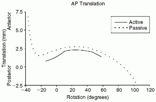
The rotator cuff and glenohumeral joint capsule function to maximize range of motion and stability in the normal shoulder. During active joint positioning within physiologic ranges of motion, the rotator cuff compresses the humeral head and dampens not only the maximum rotational motion achievable but also limits translation of the humeral head on the glenoid to 2 mm or less in most cases. The glenohumeral capsular ligaments, in their normal state, allow maximum passive range of motion and act as checkreins at the extremes of motion to prevent overrotation. In the absence of the stabilizing effect of the rotator cuff, tightness in the capsular ligaments results in greater
degrees of translation in the direction opposite the capsular tightening (Fig. 7-3) (22).


Pathologic Anatomy


Primary Osteoarthritis

The cardinal features of primary osteoarthritis in any joint are asymmetric joint space narrowing, subchondral sclerosis, subchondral cyst formation, and prominent osteophyte formation. Neer described the primary features of glenohumeral arthritis to be thinning of the humeral articular cartilage most pronounced in the area of contact when the arm is between 60 and 100 degrees of elevation (5). This is thought to be the area of greatest contact stresses. In addition, Neer described humeral subchondral cysts to be most often found slightly above the equator of the articular surface. The largest humeral osteophytes are found inferiorly. Moreover, the glenoid surface is most often flattened, devoid of articular cartilage, with palpable osteophytes within the attachment sites of the glenohumeral ligaments (5,23).

Glenoid retroversion is often increased in primary glenohumeral osteoarthritis (18,19,24, 25, 26). This may be the result of asymmetric posterior glenoid wear, congenital hypoplasia, or developmental abnormalities. In addition, posterior humeral subluxation can be found in up to 45% of shoulders with primary osteoarthritis (26). Walch and colleagues have described three types of glenoid shapes associated with primary osteoarthritis. These include type A-concentric, with no humeral subluxation (59%), type Basymmetric wear of the posterior glenoid rim with posterior humeral subluxation (32%), and Type C-glenoid retroversion of greater than 25 degrees with posterior humeral subluxation (9%) (Fig. 7-4) (25,26).


Figure 7-3 In a cadaver model, humeral head translations are significantly larger during passive rotation than active rotation because the force of the rotator cuff limits excursion. (From Karduna AR, Williams GR, Williams JL, et al. Kinematics of the glenohumeral joint: influences of muscle forces, ligamentous constraints, and articular geometry. J Orthop Res 1996;14(6):986-993, with permission.)


Figure 7-4 Glenoid shapes in osteoarthritis have been classified into types A (concave), B (biconcave), and C (retroversion >25 degrees). (From Walch G, Badet R, Boulahia A, et al. Morphologic study of the glenoid in primary glenohumeral osteoarthritis. J Arthroplasty 1999;14(6):756-760, with permission.)

Soft-tissue alterations in primary glenohumeral osteoarthritis include the capsular ligaments and the rotator cuff. The most common soft-tissue contracture involves the anterior capsule and, in severe cases, the subscapularis (5,26). In rare cases of severe posterior subluxation, the posterior capsule may be lax. However, in most cases of posterior subluxation, the head is displaced medially as well as posteriorly, resulting in less posterior capsular laxity than one would expect. The rotator cuff, especially the supraspinatus, may commonly have partial tearing. However, the incidence of full-thickness rotator cuff tears is low (5% to 10%) (5,6,27,28).


Avascular Necrosis

Except in the later stages of the disease, anatomical derangement in humeral AVN is mild and limited to the humeral bone. The hallmark of humeral osteonecrosis is loss of the cartilaginous and cancellous bony architecture from subchondral fracture and collapse. This results in replacement of the normally soft cancellous bone of the proximal humeral metaphysis with compact, hard, necrotic bone. This must be remembered when attempting to ream and broach the proximal humerus during humeral replacement. The glenoid is most often normal or mildly involved, except in the later stages of the disease, when the misshapen humeral head has been articulating against the glenoid for an extended period (29, 30, 31, 32, 33, 34, 35).

In the early stages of AVN, the rotator cuff and capsule are normal. Even as the disease progresses, the development of full-thickness rotator cuff tears is rare. The one exception to this is steroid-induced AVN associated with renal failure. As the humerus collapses and the joint becomes more arthritic, global capsular contracture and subsequent stiffness develop (31,35).


PATHOPHYSIOLOGY


Osteoarthritis

Primary osteoarthritis is the most common type of arthritis and is defined by slowly progressive, focal, and asymmetric loss of articular cartilage and hypertrophic reaction in the subchondral bone. Its cause is unknown, and it is irreversible. Age is the most well-recognized risk factor for primary degenerative disease of the shoulder. A genetic predisposition is strongly suspected (36, 37, 38), but specific chromosomal loci have yet to be identified.

The precise pathogenesis of primary osteoarthritis of the shoulder, like primary osteoarthritis anywhere in the body, is unknown. When searching for causes of osteoarthritis, two major hypotheses or models surface (39,40). One model emphasizes the role of physical forces and their ability to cause failure of articular cartilage. The other model points toward a failure of the regulation between cartilage degradation and repair. The common theme between these two theories is eventual cartilage breakdown. Other well-documented factors that have been associated with the development of osteoarthritis include aging, trauma, and heredity.

Both of the previously mentioned hypotheses or models will have the same outcome: osteoarthritis. The differences lie in the initiating events. In the first model, physical forces are responsible for articular cartilage failure. This failure could come from normal wear over a long period (increased incidence of osteoarthritis with aging) or abnormally excessive wear over a shorter period (trauma or instabilityinduced osteoarthritis). In the second model of impaired cartilage regulation, there may be an age-related decline in cartilage metabolic activity leading to inadequate maintenance (i.e., increased degradation relative to repair) of the cartilage and increased susceptibility to damage.

Once the articular cartilage is damaged, the ultimate pathway is similar no matter what the inciting event. Initially, a mild synovitis develops and an attempted repair process is begun. As a result, there is a release of degradative enzymes such as collagenase, gelatinase, and stromelysin, and a variety of inflammatory mediators, which further damage the cartilage and eventually the underlying bone (40).

The incidence of degenerative arthritis increases with age, the most common presentation occurring in the sixth decade. Primary osteoarthritis is much more prevalent in weightbearing joints of the lower extremity than it is in the shoulder. Difficulty in establishing an early diagnosis, timing of onset, and the absence of longitudinal data make estimation of its incidence and prevalence imprecise (41). However, osteoarthritis remains the primary indication for humeral head replacement. Currently, up to 60% of all shoulder arthroplasty are performed for primary osteoarthritis (5,42, 43, 44, 45).


Avascular Necrosis

Osteonecrosis is also known by the terms avascular necrosis or aseptic necrosis. At its most basic level, the process involves death of bone, both osteocytes, and marrow components (46). As with osteoarthritis, osteonecrosis is further defined as primary or secondary. Primary disease is idiopathic, and no associated cause, systemic or traumatic, can be attributed to its development. As our understanding of the pathogenesis of AVN grows, many cases previously regarded as idiopathic are indeed secondary to a known risk factor. Secondary osteonecrosis can be further broken down into traumatic and nontraumatic. The discussion in this chapter is limited to idiopathic and nontraumatic AVN, because traumatic injuries are covered elsewhere in this text.

The essential feature of osteonecrosis is the death of bone. Much of the literature supports a disruption of vasculature as the primary etiologic event. In traumatic injuries, this vascular interruption is easily understood as the mechanical disruption of the primary tributaries to the humeral head. Osteonecrosis related to a systemic etiology is less well understood but likely is related to thrombotic/embolic events or results from compression from edema or other source of extrinsic pressure (47). The etiologies of aseptic necrosis have been categorized pathologically by Mankin and colleagues as (a) mechanical vascular interruption, (b) thrombosis and embolism, (c) injury to or pressure on a vessel wall, and (d) venous occlusion (48).

Although our understanding of the pathophysiologic details is incomplete, many risk factors have been linked to the development of AVN (Table 7-1). Systemic corticosteroid use has been shown by Hattrup and Cofield to be the leading cause of nontraumatic osteonecrosis, accounting for 56% of cases (49,50). Among the other common associations are sickle cell disease and other hemoglobinopathies, Gaucher’s disease, dysbarism (caisson disease), and alcohol abuse.

The progression of pathologic changes in osteonecrosis is best described by Cruess(51), who developed a classification system based on that of Ficat for the same disease process in the hip (Table 7-2) (52). This system is based on radiographic appearance and carries both management and prognostic significance. Stage I is detectable only by magnetic resonance imaging (MRI) or bone scanning and represents bony edema and early histopathologic evidence of cell death. Stage II osteonecrosis is characterized by bony remodeling and sclerosis. In Stage III, subchondral fracture is evident, but the articular contour remains intact. A “crescent sign” is the radiographic marker of Stage III disease. Stage IV osteonecrosis is characterized by subchondral collapse and flattening of the articular contour. Advanced osteonecrosis (Stage V) occurs when degenerative change involves the glenoid as a result of the progressive articular incongruity(47) (Fig. 7-5).


TABLE 7-1 ETIOLOGY OF OSTEONECROSIS


Relationship Clearly Established


Relationship Probable


Traumatic


Traumatic


Proximal humeral fractures


Repetitive trauma


Chronic shoulder dislocations


Nontraumatic


Nontraumatic


Steroids


Dysbaric disorders


Alcohol ingestion


Gaucher’s disease


Cushing’s disease


Sickle cell anemia


Lipid metabolism disorders


Radiation


Fatty liver


Pancreatitis


Renal transplant


Systematic lupus


Erythematosus


Osteomalacia


Lymphoma


Cytotoxic drugs


Thrombophilic or hypofibrinolytic coagulopathies


From Collins DN. Pathophysiology, classification and pathoanatomy of glenohumeral arthritis and related disorders. In: Iannotti JP, Williams GR Jr, eds. Disorders of the shoulder: Diagnosis and management.


Philadelphia: Lippincott Williams & Wilkins, 1999.


TABLE 7-2 FICAT CLASSIFICATION OF OSTEONECROSIS OF THE HUMERAL HEAD


Class


Normal x-ray


I


Magnetic resonance imaging shows localized bone edema


Increased uptake on bone scan


II


Focal sclerosis in superocentral humeral head without collapse


III


Subchondral collapse “Crescent sign”


Articular surface remains intact


IV


Displaced cartilaginous flap


Articular incongruity


Degenerative change progresses to involve larger portion of humeral surface


V


Degenerative disease involves glenoid


The osteonecrotic lesion is typically located at the site of glenohumeral contact when the shoulder is in approximately 90 degrees of shoulder abduction (29,32,51). Biomechanical studies have shown this to be the point of maximal force transmission across this articulation (53).

The overall incidence of posttraumatic osteonecrosis is difficult to accurately determine because many of these patients do not require medical attention beyond the initial diagnostic visit and are satisfied with conservative management. The majority of these patients do not require surgical intervention for symptomatic control (54). Cases of posttraumatic osteonecrosis far outnumber idiopathic cases and aseptic necrosis related to a systemic etiology. The humeral head is the second most common site for nontraumatic osteonecrosis after the femoral head; osteonecrosis is the indication for shoulder arthroplasty in approximately 3% to 5% of cases (55).


Figure 7-5 Humeral osteonecrosis can be staged as I—bone edema and early histologic signs of cell death, II—bone remodeling and sclerosis, III—subchondral fracture (i.e., crescent sign) with intact articular contour, IV—subchondral collapse and articular flattening, and V—glenoid involvement. (Modified from Cofield RH. Osteonecrosis In: Friedman RJ, ed. Arthroplasty of the Shoulder. New York: Thieme, 1994;174, with permission.)

As previously discussed, because of its position as a nonweightbearing joint, the shoulder has a more favorable prognosis with AVN than the hip. The rate and degree of progression for an individual is not predictable, although some trends have been noted (54,56). Reports indicate that in general, traumatic osteonecrosis has a more rapidly progressive course than nontraumatic cases. In a group of patients followed for 3 years after their diagnosis, only 43% of patients with corticosteroid-induced osteonecrosis required arthroplasty, versus 80% of posttraumatic cases (54). The radiographic stage at diagnosis has also been shown to positively correlate with rate of disease progression (57). Patients with sickle cell disease tend to have the lowest rate of arthroplasty among the various causes of nontraumatic osteonecrosis. In a series of 2,500 patients with radiographic evidence of humeral head osteonecrosis, 21% of whom had pain and stiffness at presentation, only one patient went on to require arthroplasty (56). Another study reported that only 2 of 138 patients with sickle cell disease in their series ultimately required arthroplasty (35).


EVALUATION


History

Patients with primary osteoarthritis typically present with an insidious onset of pain, which has been slowly progressive over past years. Progressive stiffness is often associated with the discomfort. As is the case in other arthritic joints, the pain is often activity related. Stiffness is most notable after a period of immobilization and improves with gentle motion.

Patient complaints will often relate to functional limitations such as difficulty internally rotating to reach their back pocket or fastening a brassiere. Questioning the patient as to how their symptoms have interfered with their daily routines will provide insight into the degree of pain and disability. Understanding the patient’s occupation, hobbies, and activity levels also helps gauge the impact of their disease. Documentation of treatments that have been or are being used also gives information about disease course and severity. Awareness of the patient’s profile of comorbidities is important not only for presurgical screening but also as a means of evaluating what other conditions might limit activity and rehabilitation.

Patients suspected to have AVN should also be questioned regarding the onset, course, severity, and impact of their symptoms because this is valuable information in management decision making. Typically, these patients are younger than those presenting with osteoarthritis. Identification of risk factors can aid in determining the etiology of osteonecrosis. Exposure to steroids, alcohol use or abuse, liver pathology, and personal or family history of coagulation disorders should be addressed. Careful questioning about pain in the contralateral shoulder and other joints at risk (hips, knees, ankles, etc.) is also important. Although only 3% of patients with osteonecrosis have multifocal involvement (58,59), 80% of patients with multifocal osteonecrosis will have humeral head involvement (58).


Physical Examination

A complete shoulder examination should be performed with particular attention to range of motion. Typically both osteoarthritis and AVN cause progressive global loss of motion, with particular loss of external rotation. Any internal rotation contracture must be noted and documented because it dictates whether subscapularis releases are required at the time of surgery. Active and passive motion should be compared, and the rotator cuff strength should be noted. This can sometimes be difficult to determine on physical examination alone because there is often pain-related weakness. Painful crepitus is common. Tenderness to palpation over the acromioclavicular joint can indicate arthritis, which potentially can contribute to the symptom complex. This is an important finding to identify because it can also be addressed at the time of surgery if needed.


Imaging Studies

Plain radiographs are the single most important investigation required in the diagnosis of osteoarthritis and arthroplasty planning. A standard x-ray series (anteroposterior [AP], transcapular lateral, and axillary lateral) usually is performed, and each provides different information required for preoperative preparation. The AP x-ray, which often is done in internal and external rotation, allows assessment of bone quality, identification of inferior osteophytes, and diameter of the humeral canal. Preoperative templating is performed using the AP and axillary views. Also, although not universally reliable because of slight variations in angle of beam projection, evaluation of the acromiohumeral distance can suggest the presence of significant rotator cuff deficiency if the distance measures less than 7 mm. The axillary radiograph is useful in identifying glenoid version and the posterior glenoid wear and resultant posterior subluxation that is often associated with osteoarthritis (Fig. 7-6).

CT scan provides a more definitive assessment of glenoid bone stock and version. It also allows accurate determination of whether glenoid replacement is feasible and if bone grafting may be necessary (Fig. 7-7). One radiographic study of a series of patients with primary glenohumeral osteoarthritis who were awaiting shoulder arthroplasty found that 45% have posterior subluxation (26). Average glenoid retroversion in this population was found to be 15.4 degrees (normal being 1 to 2 degrees of retroversion) (19). Humeral version can also be accurately determined from CT pictures of the humeral head and its relation to the transcondylar axis (60). Because posterior glenoid erosion and posterior subluxation are common with severe internal
rotation contracture, CT scanning is ordered in all patients with external rotation of 30 degrees or less.

Only gold members can continue reading. Log In or Register to continue

Stay updated, free articles. Join our Telegram channel

Sep 16, 2016 | Posted by in ORTHOPEDIC | Comments Off on Replacement Arthroplasty in Glenohumeral Arthritis: Intact or Reparable Rotator Cuff

Full access? Get Clinical Tree

Get Clinical Tree app for offline access