Recent guidelines have suggested that routine postoperative care of patients with metal-on-metal hip prostheses should involve metal ion analysis. This study sought to investigate the relationship between bearing surface wear rates of metal components and serum metal ion analysis and also to quantify the incidence of excessive increases in serum metal ion concentrations post–hip resurfacing arthroplasty.
Increased chromium (Cr) and cobalt (Co) concentrations following metal-on-metal (MoM) hip arthroplasty are associated with local and systemic pathologic changes. It is unquestionable that efforts should be made to identify modifiable variables leading to ion release to minimize the risk of adverse effects.
To date, the variables that have been shown to significantly affect metal ion concentrations in the absence of renal disease are femoral component diameter, acetabular cup angles of inclination and anteversion, time from surgery, and activity. Of these variables, only cup orientation can be regarded as realistically under the surgeon’s control. However, it remains to be proven whether an optimal cup position exists and whether this is affected by implant design. Our previous work and those of others have suggested that the coverage angle provided by the acetabular cup is critical. There is evidence, concordant with laboratory data, to suggest that clearance may also be an important variable in determining in vivo wear rates.
There were 3 aims of this study:
- 1.
To investigate the relationship between volumetric wear rate and serum metal ion concentrations
- 2.
To establish the incidence of excessive metal ion release following resurfacing with commonly used devices
- 3.
To identify cup orientations associated with lowest ion concentrations for each device and to propose mechanisms leading to increased wear.
Patients and methods
There were 723 patients in this series, 446 men and 277 women. All surgeries were performed by 2 experienced hip resurfacing surgeons who use metal ion analysis as part of routine follow-up. Both surgeons perform more than 100 hip resurfacings per year. Surgeon 1 is based in the United Kingdom (at site 1) and surgeon 2 is based in Belgium (at site 2). At both sites, blood samples were collected from the patients at a minimum time of 12 months after surgery to avoid the confounding factor of higher levels of wear during the run-in period. These patients did not have any other metal implants at the time of measurement. Three resurfacing devices were studied: the Articular Surface Replacement (ASR; DePuy International, Leeds, UK), the Birmingham Hip Resurfacing (BHR; Smith and Nephew, Warwick, UK), and the Conserve® Plus (C+; Wright Medical Technology, Memphis, TN, USA). Serum ion analysis was performed using inductively coupled plasma mass spectrometry. Volumetric wear rates of explanted components determined by coordinate measuring machine analysis were compared with corresponding serum ion concentrations in vivo. The relationships between ion levels and component size and cup orientations remaining in vivo were investigated. The mean (range) age of patients from site 1 was 56 years (25–83 years) and the mean (range) time from surgery to blood sampling was 29 months (12–72 months) for the patients with the ASR implant device and 66 months (48–81 months) for those with the BHR implant device. The mean age of patients from site 2 was 51 years and the mean (range) time from surgery to blood sampling was 19 months (12–29 months) for the patients with the ASR implant device, 51 months (12–106 months) for those with the BHR implant device, and 28 months (12–54 months) for those with the C+ implant device.
Implants
From site 1, there were 223 patients with ASR and 72 with BHR implants. From site 2 there were 271 patients with BHR, 136 with C+, and 21 with ASR implants. Table 1 shows the differences in design between the implants in this study.
| Device Characteristics | ASR | BHR | C+ |
|---|---|---|---|
| Subtended articular surface angle (°) | 144–160 a | 158–165 a | 162–165 a |
| Mean nominal radial clearance (μm) | 50 | 100 | 80 |
| Manufacturing method of head | As cast | As cast | HIP/SA b |
| Manufacturing method and treatment of cup | HIP/SA b | As cast | HIP/SA b |
| Carbon content c | High | High | High |
a Subtended articular surface angles increase with increasing cup diameter.
b HIP/SA, cast process and heat treatment by hot isostatic pressure/surface annealed.
Sample Collection
The samples were obtained at site 2 using an intravenous catheter (Insyte-WTM; Becton Dickinson, Franklin Lakes, NJ, USA). After the catheter had been introduced, the metal needle was withdrawn and the first 5 mL of blood was discarded to avoid possible contamination from the needle. A second 5 mL was collected using a vacuum tube (Venosafe VF-106SAHL; Terumo Europe NV, Leuven, Belgium). The samples were analyzed at the Laboratory of Clinical Biology at Ghent University Hospital, Ghent, Belgium. The laboratory quotes its quantification limit as 0.5 μg/L with a reproducibility of 5%. At site 1, serum samples were collected in a similar manner. Venous cannulation was performed with a 21-gauge stainless steel needle (Venflon, Becton Dickinson, Helsingborg, Sweden), with disposal of the first 5 mL of blood to avoid contamination. All samples were centrifuged to separate blood and serum fractions, frozen, and sent for blinded trace element analysis at the Trace Element Laboratory of the Royal Surrey County Hospital, Guildford, United Kingdom. This laboratory also quotes its quantification limit as 0.5 μg/L with a reproducibility of 5%.
Radiographic Analysis
At the time of collection of the sample, the University of California, Los Angeles activity scores were recorded and weight-bearing pelvic radiographs were obtained. From these radiographs, the following parameters were measured using ImageJ software (National Institutes of Health, Bethesda, MD, USA): femoral stem angle relative to the femoral shaft (SSA), femoral stem to femoral neck angle (SNA), and femoral offset and femoral component to femoral neck ratios. Einzel-Bild-Roentgen-Analyse (EBRA, University of Innsbruck, Innsbruck, Austria) software was used to analyze all available radiographs to obtain angles of cup inclination and anteversion. The accuracy of this software has been discussed in the literature. In most cases, one well-centered radiograph was available for analysis. Theoretical contact patch to rim (CPR) distance was calculated for all patients.
To test the validity of comparing Cr and Co levels between the 2 populations, patients with BHR implants from site 2 were matched with patients with BHR implants from site 1 by the length of time to blood sampling from surgery. From these patients, we selected only those with femoral components of size 50 mm and larger as we have previously showed these sizes to be relatively resistant to cup position. To further control for the effects of cup position, cups with inclinations greater than 55° and less than 10° or anteversion greater than 30° were excluded. The Cr and Co levels for the matched populations were then compared using Mann-Whitney tests for nonparametric data ( Table 2 ). P values less than .05 were deemed significant. Two sample t -tests comparing the relevant parameters can be seen in Table 2 . Cr concentrations in patients from UK were found to be slightly but significantly increased when compared with patients from Belgium. There was no significant difference between the groups with regard to Co. For this reason, we used only serum Co in the analysis.
| Variables | Site 1 | Site 2 | P value |
|---|---|---|---|
| Number | 40 | 48 | — |
| Time to Sample (mo) | 66 (48–81) | 64 (50–81) | .190 |
| Femoral Size (mm) | 51.6 | 52.4 | .107 |
| Cup Inclination (°) | 48.5 (33–55) | 48.8 (34–55) | .890 |
| Cup Anteversion (°) | 17.04 (10–39) | 20.5 (10–36) | <.001 |
| Serum Cr (μg/L) a | 3.49 | 1.80 | <.001 |
| Serum Co (μg/L) a | 1.31 | 1.20 | .812 |
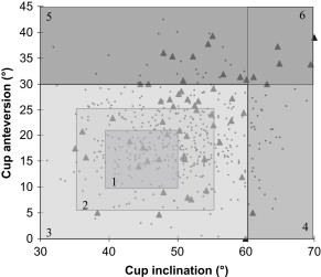
For the 3 resurfacing devices, the relationship between each variable and serum Co was examined using the Spearman rank correlation. Rank correlation was preferred because of the nonparametric nature of the ion data. Scatter plots were used to identify nonlinear relationships. To examine the relative sensitivity of each device to the effects of cup orientation, the patients from each implant group were divided into subgroups of inclination (<25°, 25°–27°, 27°–29°, 29°–31°, 31°–33°, 33°–35°) and anteversion (<0°, 0°–5°, 5°–7°, 7°–9°, 9°–11°). From each subgroup the median Co level was calculated and plotted. To further examine this relationship, patients were placed into zones of cup orientation based on the EBRA measurements ( Fig. 1 ). We then calculated the percentage of patients in each zone (for each device) with serum Co levels more than 7 μg/L. This figure has recently been quoted by the Medicines and Healthcare Products Regulatory Agency/Metal on Metal Working Committee of the United Kingdom, indicating a poorly performing bearing surface with recommendations for close patient follow-up. Windows SPSS version 15.0 (SPSS Inc, Chicago, IL, USA) was used for statistical analysis throughout.
Survival Analysis Using Metal Ion Concentrations
In our previous work we showed that there was a suggestion that there is a temporary immunity to increases in the wear of malpositioned cups/cups. With this principle in mind, we collected all the metal ion results from all patients in the study at all periods, including repeat samples from individual patients at different times postoperatively. Of the 723 patients, 124 had undergone repeat testing (as part of routine screening), giving a total of 847 samples. Using these data, we conducted a Kaplan-Meier survival analysis comparison between the patients in each device group, with the latest follow-up being the time in months from the resurfacing to the last blood sample. The patients were then categorized into a large joint group (femoral diameter≥53 mm) or a small joint group (femoral diameter<53 mm). The principle of this categorization was based on our previous results, which showed larger joints to be less vulnerable to the effects of cup position. The patients were then also categorized by cup position and a Cox proportional hazard model constructed.
Explant analysis
The wear of retrieved ASR matched femoral and acetabular components from site 1 (n = 35 pairs) was measured by a coordinate measuring machine (CMM) using a scanning head (Legex 322, Mitutoyo Halifax, UK) with a spatial resolution of less than 1 μm in the area of measurement. Measurements were made for every 5° on 18 concentric circles as well as at the pole of the component, which resulted in a total of 4500 to 6000 measurements for each component, dependent on the radius of the explant. The wear scars of each component were categorized as anteverted or retroverted according to the description by Walter and colleagues. Volumetric wear was calculated using the method described in the Appendix . Univariate linear regression was used to examine the relationship between volumetric wear rates and serum Co and Cr levels. Using the Medicines and Healthcare products Regulatory Agency (MHRA) level of 7 μg/L as a positive test and abnormal bearing surface wear as an annual wear rate greater than 3 mm 3 /y, (In our recent work, 3 mm 3 /yr was the lowest bearing surface wear rate of a resurfacing device associated with adverse reaction to metal debris. ) the sensitivity and specificity of serum Cr and Co testing was evaluated.
Results A: Ex vivo
Explant Analysis and Serum Ion Concentrations
Volumetric wear correlated well with serum Cr (r 2 = 0.66, P <.01) and serum Co concentrations (r 2 = 0.78, P <.01). Specificity and sensitivity (95% CI) of serum Co levels greater than 7 μg/L were found to be 1.00 (0.67–1.00) and 0.93 (0.83–0.98), respectively. Specificity and sensitivity (95% CI) of serum Cr levels greater than 7 μg/L were found to be 0.80 (0.48–0.97) and 0.93 (0.83–0.97), respectively. Wear maps of retrieved components were generated and can be seen adjacent to the corresponding radiographs ( Fig. 2 ). In Fig. 2 , for simplification, red areas represent wear of greater than 20 μm deviation from a perfect spherical form. All explants that were found to have extremely high wear rates were found to have edge loading of the acetabular components. We define edge loading here as a progressive increase in the measured wear depths of the cups so that maximal wear depths occur at the edge of the articular surface.
Results B: In vivo
Femoral Size
In all 3 devices, we identified an inverse relationship between femoral component size and serum Co concentrations ( Table 3 ). This relationship was not significant in the C+ device. However, the largest values were found in the patients in whom larger-diameter bearings had been suboptimally positioned.
Related posts:
 Complications After Metal-on-Metal Hip Resurfacing Arthroplasty
Imaging of Metal-On-Metal Hip Resurfacing
The Effect of Patient Selection and Surgical Technique on the Results of Conserve® Plus Hip Resurfacing—3.5- to 14-Year Follow-up
Sporting Activity After Hip Resurfacing: Changes Over Time
Incidence and Significance of Femoral Neck Narrowing in the First 500 Conserve® Plus Series of Hip Resurfacing Cases: A Clinical and Histologic Study
The Future of Hip Resurfacing
Complications After Metal-on-Metal Hip Resurfacing Arthroplasty
Imaging of Metal-On-Metal Hip Resurfacing
The Effect of Patient Selection and Surgical Technique on the Results of Conserve® Plus Hip Resurfacing—3.5- to 14-Year Follow-up
Sporting Activity After Hip Resurfacing: Changes Over Time
Incidence and Significance of Femoral Neck Narrowing in the First 500 Conserve® Plus Series of Hip Resurfacing Cases: A Clinical and Histologic Study
The Future of Hip Resurfacing
Stay updated, free articles. Join our Telegram channel

Full access? Get Clinical Tree


