 Plantar Approach to the Lateral Sesamoid Bone
The plantar approach to the lateral sesamoid bone is used exclusively for excision of the lateral sesamoid. As with all plantar approaches through areas of weight-bearing skin, there is the possibility of creating an uncomfortable scar. The common digital nerve supplying the skin of the first web space is at risk during this approach.
Peripheral vascular disease with absent peripheral pulses is a major contraindication to this approach. Delayed wound healing or necrosis may occur if the vascular supply is compromised. Preoperatively, a careful, systematic examination of the vascular supply to the foot is mandatory.
Position of the Patient
Place the patient supine on the operating table. After exsanguination, use a tourniquet placed on the middle of the thigh. Alternatively, use a soft rubber bandage to exsanguinate the foot, then wrap the leg tightly just above the ankle (see Fig. 1-1).
Landmarks and Incision
Palpate the heads of the first and second metatarsal bones on the plantar aspect of the foot. Passive flexion and extension of the hallux and index toe will allow you to identify the level of the metatarsophalangeal joint.
The sesamoid bones themselves may be palpable. They can be felt by applying pressure with the thumb to compress them against the underlying first metatarsal head.
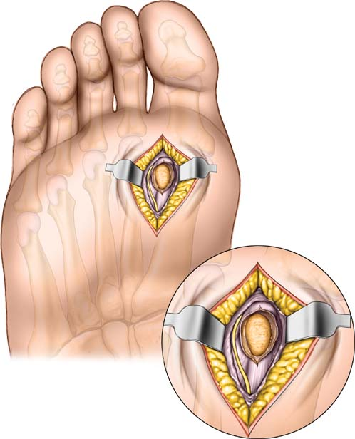
Make a 4-cm longitudinal incision on the plantar aspect of the foot between the first and second metatarsal heads. Begin the incision at the level of the metatarsophalangeal joint of the hallux and proceed proximally. This skin incision passes lateral to the lateral sesamoid bone (Fig. 40-1).
Related posts:

Stay updated, free articles. Join our Telegram channel

Full access? Get Clinical Tree


