Pictorial Guide to Muscles and Surface Anatomy


Pictorial Guide to Muscles and Surface Anatomy


Henry L. Lew

Su-Ju Tsai


Table 8-1 Masseter (Fig. 8-1)


image
Figure 8-1 • Masseter muscle
Innervation Mandibular division of the trigeminal nerve (cranial nerve V), mesencephalon
Origin Lower and medial aspect of the zygomatic arch
Insertion Lateral aspect of the mandibular ramus
Action Elevation of the mandible for mastication
Position Supine with the head turned to the other side or lateral decubitus
Activation Teeth clenching
Needle placement This muscle is midway between the angle of the jaw and the temporomandibular joint.
Notes

  1. Needle is held at oblique angle of 20 degrees from the skin. Muscles are too thin for perpendicular insertion.
  2. Motor unit potentials are smaller in both amplitude and duration than those seen in limb muscles.


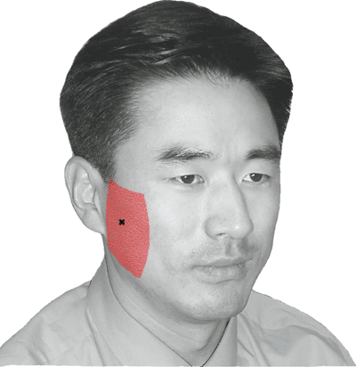
Table 8-2 Temporalis (Fig. 8-2)


image
Figure 8-2 • Temporalis
Innervation Deep temporal nerve—mandibular division of the trigeminal nerve (cranial nerve V), mesencephalon
Origin Bony floor of the temporal fossa and the deep surface of the temporal fascia
Insertion Coronoid process and anterior border of the ramus of the mandible
Action Elevation and retraction of the mandible
Position Supine with the head turned to the other side or lateral decubitus
Activation Teeth clenching
Needle placement Vertically above its insertion at the mandible, insert 1–2 cm below the skull ridge from which it arises. Palpate temporal artery to avoid.
Notes See notes in Table 8-1.


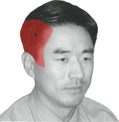
Table 8-3 Auricularis Posterior (Fig. 8-3)


image
Figure 8-3 • Auricularis posterior
Innervation Facial nerve (cranial nerve VII), pons
Origin Mastoid process
Insertion Cartilage of the ear
Action Retraction of the earlobe
Position Supine with the head turned to the other side or lateral decubitus
Needle placemen Posterior to the midpoint of the ear; can be absent and some patients cannot activate voluntarily
Notes See notes in Table 8-1.


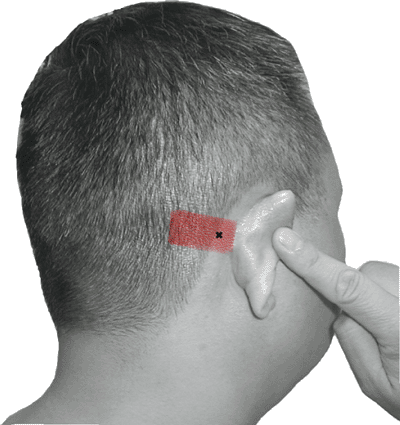
Table 8-4 Frontalis (Fig. 8-4)


image
Figure 8-4 • Frontalis
Innervation Temporal branch of the facial nerve (cranial nerve VII), pons
Origin Frontal skin and superficial fascia of the eyebrow
Insertion Epicranial aponeurosis
Action Raise the eyebrows
Position Supine
Activation Raise the eyebrows or look upward.
Needle placement The frontalis muscle is 5 cm above the eyebrow, superior to most of the wrinkles on the forehead that it makes.
Notes See notes in Table 8-1.


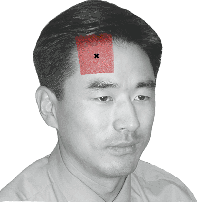
Table 8-5 Orbicularis Oculi (Fig. 8-5)


image
Figure 8-5 • Orbicularis oculi
Innervation Temporal and zygomatic branches of the facial nerve (cranial nerve VII), pons
Origin Medial palpebral ligament and adjacent bones
Insertion Lateral palpebral raphe and adjacent tissues
Action Closure of the eyelids and dilatation of the lacrimal sac
Position Supine
Activation Close eyes, resist opening.
Needle placement Palpate the margin of the orbital fossa, and insert the needle in 20° lateral to the margin. Direct the needle to avoid the eyeball.
Notes See notes in Table 8-1.


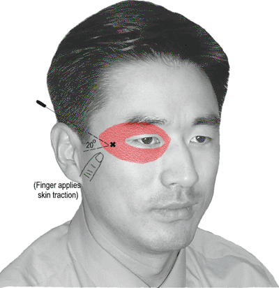
Table 8-6 Nasalis (Fig. 8-6)


image
Figure 8-6 • Nasalis
Innervation Buccal branch of the facial nerve (cranial nerve VII), pons
Origin Maxilla
Insertion Ala of the nose
Action Widening of the nasal aperture
Position Supine
Activation Deep inspiration through the nostril
Needle placement Study this muscle just below the bone–cartilage junction of the nose.
Notes See notes in Table 8-1.


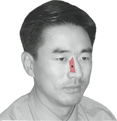
Table 8-7 Mentalis (Fig. 8-7)


image
Figure 8-7 • Mentalis
Innervation Mandibular branch of the facial nerve (cranial nerve VII), pons
Origin Mandible
Insertion Skin on the chin
Action Protrusion of the lower lip
Position Supine
Activation Protrude the lower lip; “pucker.”
Needle placement Just above the corners of the anterior jaw
Notes See notes in Table 8-1.


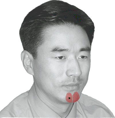
Table 8-8 Orbicularis Oris (Fig. 8-8)


image
Figure 8-8 • Orbicularis oris
Innervation Buccal and mandibular branches of the facial nerve (cranial nerve VII), pons
Origin Encircling fibers from the deep surface of the skin, the buccinator
Insertion Skin and mucous membrane lining the inner surface of the lips
Action Compressing the lips together
Position Supine
Activation Lip pucker
Needle placement This is the deepest-lying of the facial muscles; insert just lateral to mouth angle, with 30° angle for deeper penetration.
Note See notes in Table 8-1.


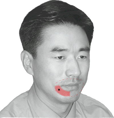
Table 8-9 Sternocleidomastoid (Fig. 8-9)


image
Figure 8-9 • Sternocleidomastoid
Innervation Spinal accessory nerve (cranial nerve XI) and the anterior rami of the C2 and C3 nerves, medulla
Origin Upper portion of the sternum and the medial third of the upper surface of the clavicle
Insertion Mastoid process and lateral portion of the superior nuchal line of the occipital bone
Action Tilting of the head to the same side, and turning the chin to the opposite side
Position Supine
Activation Rotate the head to opposite side.
Needle placement Midway between the mastoid and the clavicle; pinch muscle and lift to avoid penetrating too deeply.


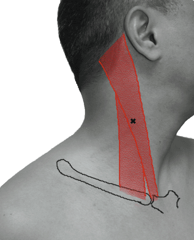
Table 8-10 Trapezius (Fig. 8-10)


image
Figure 8-10 • Trapezius
Innervation Spinal accessory nerve (cranial nerve XI), C3 and C4 cervical nerves, medulla
Origin External occipital protuberance, ligamentum nuchae, and spinous processes of the C7 and T1–T12 vertebrae
Insertion Lateral third of the clavicle, the acromion, and the spine of the scapula
Action Adduction and rotation (elevating the acromion) of the scapula
Position Prone, lateral decubitus, or sitting
Activation Shrug shoulders.
Needle placement Draw a line from the C7 spinous process to the acromion and insert in the center of this line.


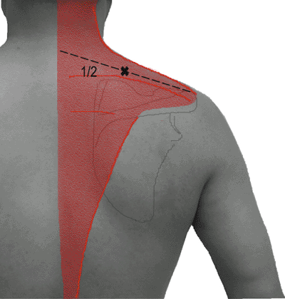
Table 8-11 Tongue (Fig. 8-11)


image
Figure 8-11 • Tongue
Innervation Hypoglossal nerve (cranial nerve XII), medulla
Origin Superior genial spine of the mandible
Insertion Inferior portion of the tongue blending with other tongue muscles
Action Protrusion and lateral movement of apex of the tongue
Position Supine with the head turned to the other side
Activation Tongue protrusion or contralateral movement
Needle placement Insert vertically upward 1 cm posterior to the (posterior) edge of the mental portion of the mandible and 1 cm from center.


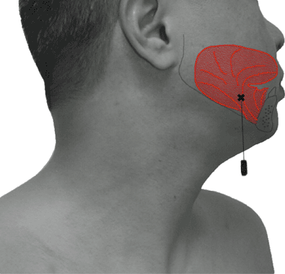
Table 8-12 Diaphragm (Fig. 8-12)


image
Figure 8-12 • Diaphragm
Innervation Phrenic nerve: C3, C4, C5
Origin Lower six ribs, internal surface of xiphoid process, medial and lateral arcuate ligaments from transverse processes of L1 and L2 vertebrae
Insertion Central tendon of the diaphragm
Action Inspiration (depression of the diaphragm)
Position Supine
Activation Rapid inspiration with pursed lips for resistance
Needle placement From the costal margin, palpate the intercostal space below the 9th rib. Insert the needle perpendicular to the skin 1 cm lateral to the anterior articulation and close to the superior edge of the 10th rib. Keep amplifier on and observe the intercostal muscle EMG activity and the respiratory pattern. Penetrate deeper with the needle reaching the diaphragm, which is active only on inspiration.
Note: For COPD patients with a depressed diaphragm at rest, insert the needle below the costal margin in the angle between the xiphoid process and the rib.


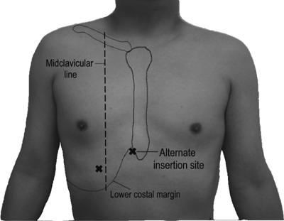
Table 8-13 Levator Scapulae (Fig. 8-13)


image
Figure 8-13 • Levator scapulae
Innervation Dorsal scapular nerve, branches from C3 and C4
Origin Transverse processes of atlas, axis, C3, and C4
Insertion Medial border of the scapula, superior to the root of the spine
Action Elevation of the scapula
Position Prone, lateral decubitus, or sitting
Activation Shrug shoulder.
Needle placement Insert into the muscle above the medial-superior aspect of the scapula and activate with elevation of the shoulder.
Note When activating muscles for motor unit evaluation, use isometric contraction.


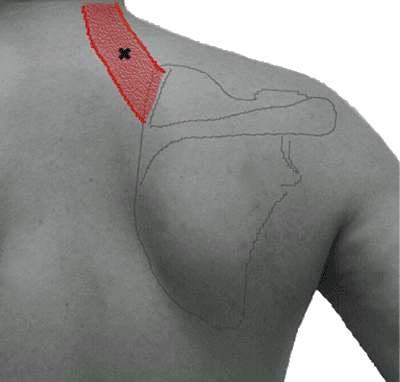
Table 8-14 Rhomboid Major (Fig. 8-14)


image
Figure 8-14 • Rhomboid major
Innervation Dorsal scapular nerve—C5
Origin Spinous processes of the T2–T5 vertebrae
Insertion Medial border of the scapula, inferior to the spine
Action Adduction, retraction, and rotation of the scapula (acromion down)
Position Prone, lateral decubitus, or sitting
Activation Retraction (adduction) of the scapula
Needle placement Just medial to the inferior third of the scapula; this is a “keep-amplifier-on” muscle studied with retraction of the scapula.
Note “Keep-amplifier-on” refers to switching on the preamplifier with the needle tip in the dermis or subcutaneous layers. The tip is slowly advanced until the needle enters the muscle. This avoids accidental penetration through the intercostal space.


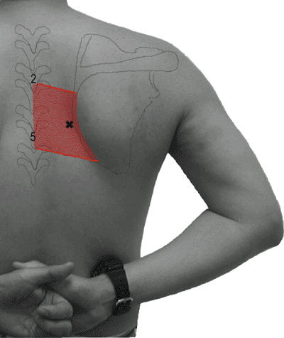
Table 8-15 Rhomboid Minor (Fig. 8-15)


image
Figure 8-15 • Rhomboid minor
Innervation Dorsal scapular nerve—C5
Origin Spinous processes of the C7 vertebra and the T1 vertebra
Insertion Medial border of the scapula at the base of the spine of the scapula
Action Adduction, retraction, and elevation of the scapula
Position Prone, lateral decubitus, or sitting
Activation Retraction of the scapula
Needle placement Immediately medial to the scapular spine, this is studied with retraction-elevation of the scapula as a “keep-amplifier-on” muscle.
Note See notes in Table 8-14.


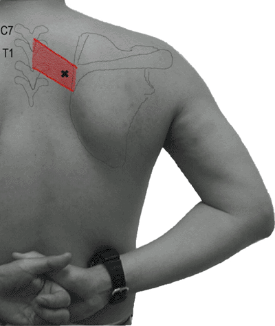
Table 8-16 Serratus Anterior (Fig. 8-16)


image
Figure 8-16 • Serratus anterior
Innervation Long thoracic nerve—C5, C6, C7
Origin Anterior and upper surfaces of the upper eight to nine ribs
Insertion Anterior surface of the medial border of the scapula
Action Abduction and protraction of the scapula
Position Lateral decubitus or supine
Activation Forward flexion or protraction of shoulder
Needle placement Palpating ribs 6 and 7 in the mid-axillary line will show this muscle, which protracts the scapula. Place fingers in the intercostal spaces while you insert the needle toward the rib between the fingers.


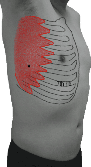
Table 8-17 Supraspinatus (Fig. 8-17)


image
Figure 8-17 • Supraspinatus
Innervation Suprascapular nerve—upper trunk—C5, C6
Origin Supraspinous fossa of the scapula
Insertion Greater tubercle of the humerus
Action Abduction and external rotation of the shoulder
Position Prone, lateral decubitus, or sitting
Activation Abduction or external rotation of the shoulder
Needle placement Just above the spine of the scapula and 2 cm from its medial border. Avoid the narrow lateral half of the scapula, which overlies apical lung tissue. By inserting the needle down to the bone, one is assured of passing through the trapezius completely.


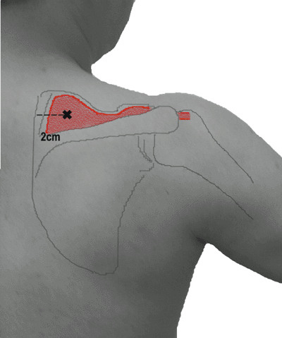
Table 8-18 Infraspinatus (Fig. 8-18)


image
Figure 8-18 • Infraspinatus
Innervation Suprascapular nerve—upper trunk—C5, C6
Origin Infraspinous fossa of scapula
Insertion Greater tubercle of the humerus
Action External rotation of the shoulder
Position Prone, lateral decubitus, or sitting
Activation Externally rotate the shoulder.
Needle placement Halfway between the inferior angle of the scapula and its spine, insert the needle 3 cm from the medial edge of the bone.


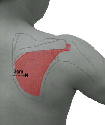
Table 8-19 Teres Major (Fig. 8-19)


image
Figure 8-19 • Teres major
Innervation Lower subscapular nerve—posterior cord—posterior division—upper trunk—C5, C6
Origin Lower third lateral border of the scapula
Insertion Medial lip of bicipital groove of the humerus
Action Adduction, extension, and internal rotation of the arm
Position Prone, lateral decubitus, or sitting
Activation Internally rotate the shoulder.
Needle placement Along the lateral scapula, 2–3 cm above the inferior angle. This is a “keep-amplifier-on” muscle so that you do not pass through it.
Note See notes in Table 8-14.


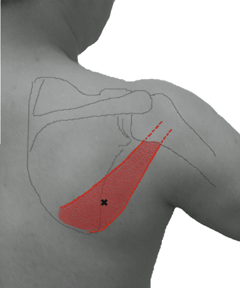
Table 8-20 Pectoralis Major—Clavicular Head (Fig. 8-20)


image
Figure 8-20 • Pectoralis major—clavicular head
Innervation Lateral pectoral nerve—lateral cord—anterior division—upper trunk—C5, C6
Origin Sternal half of the clavicle
Insertion Lateral lip of the bicipital groove of the humerus
Action Adduction, flexion, and internal rotation of the shoulder
Position Supine or sitting
Activation Internally rotate the shoulder.
Needle placement Study can be done lateral with shoulder externally rotated to extend and expose the muscle. Insert medial to the neck of the humerus. Alternatively, insert just below the middle of the clavicle.


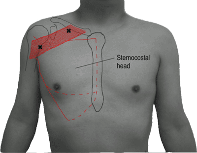
Table 8-21 Biceps Brachii (Fig. 8-21)


image
Figure 8-21 • Biceps brachii
Innervation Musculocutaneous nerve—lateral cord—anterior division—upper trunk—C5, C6
Origin Long head: supraglenoid tuberosity of the scapula
Short head: tip of coracoid process of the scapula
Insertion Bicipital tuberosity of the radius
Action Supination of the forearm and elbow flexion
Position Supination of the forearm (elbow flexion preferentially activates brachialis)
Activation Supination of the forearm
Needle placement Insert into middle of superficial muscle belly. Fully supinate the forearm to bring this muscle anterior.


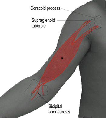
Table 8-22 Brachialis (Fig. 8-22)


image
Figure 8-22 • Brachialis
Innervation Musculocutaneous nerve—lateral cord—anterior division—upper trunk—C5, C6
Origin Lower two thirds of anterior surface of the humerus
Insertion Anterior aspect of coronoid process and tuberosity of the ulna
Action Flexion of the elbow
Position Pronation and extension of the forearm, supine
Activation Flex the elbow.
Needle placement This is the primary muscle for elbow flexion. Approach from lateral at the distal third of the arm.


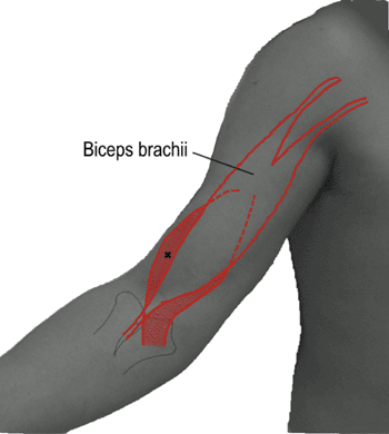
Table 8-23 Coracobrachialis (Fig. 8-23)


image
Figure 8-23 • Coracobrachialis
Innervation Musculocutaneous nerve—lateral cord—anterior division—upper and middle trunk—C6, C7
Origin Tip of the coracoid process of the scapula
Insertion Middle of medial border of the humerus
Action Flexion and adduction of the shoulder
Position Arm at side, supine
Activation Flex the shoulder.
Needle placement Insert into the anterior axillary fold. Follow muscle line out from the inferior edge of the coracoid to midhumerus.


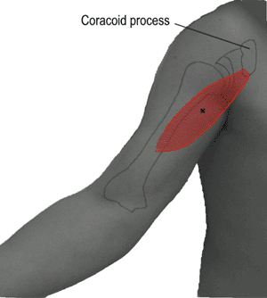
Table 8-24 Latissimus Dorsi (Fig. 8-24)


image
Figure 8-24 • Latissimus dorsi
Innervation Thoracodorsal nerve—posterior cord—posterior division—upper, middle, and lower trunk—C6, C7, C8
Origin Spinous processes of lower six thoracic vertebrae, all lumber and sacral vertebrae, and posterior iliac crest
Insertion Floor of the bicipital groove of the humerus
Action Adduction, extension, and medial rotation of the shoulder
Position Prone or lateral decubitus, full shoulder flexion
Activation Resist shoulder extension.
Needle placement The posterior axillary fold contains this larger muscle. An anterior approach helps to avoid the lower trapezius.


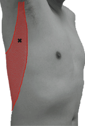
Table 8-25 Deltoid (Fig. 8-25)


image
Figure 8-25 • Deltoid
Innervation Axillary nerve (posterior and anterior branches)—posterior cord—posterior division—upper trunk—C5, C6
Origin Lateral third of the clavicle, upper aspect of acromion, and spine of the scapula
Insertion Deltoid tuberosity of the humerus
Action Abduction of the shoulder (assists with flexion and extension)
Position Anterior and middle heads: supine, lateral decubitus, or sitting
Posterior head: prone, lateral decubitus, or sitting
Activation Abduct the shoulder (flexion, anterior; extension, posterior).
Needle placement For the anterior head, insert 1 cm lateral to the coracoid process. Insert midway between acromion and the tubercle for the middle head. The posterior head is below the posterior aspect of the glenoid.
Note Posterior branch—posterior head


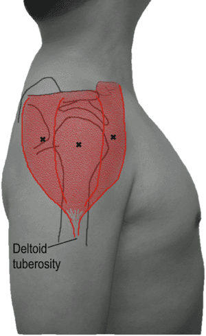
Table 8-26 Teres Minor (Fig. 8-26)


image
Figure 8-26 • Teres minor
Innervation Axillary nerve (posterior branch)—posterior cord—posterior division—upper trunk—C5, C6
Origin Upper two thirds of the lateral border of the scapula
Insertion Greater tubercle of the humerus, capsule of the shoulder joint
Action Adduction and external rotation of the shoulder
Position Prone, lateral decubitus, or sitting
Activation Externally rotate the shoulder.
Needle placement This muscle overlies the upper two thirds of the lateral border of the scapula and the lower portion of the glenohumeral joint.


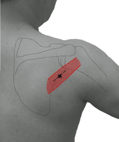
Table 8-27 Brachioradialis (Fig. 8-27)


image
Figure 8-27 • Brachioradialis
Innervation Radial nerve—posterior cord—posterior division—upper trunk—C5, C6
Origin Proximal two thirds of lateral supracondylar ridge of the humerus
Insertion Base of styloid process of the radius
Action Flexion of the forearm elbow
Position Half-pronation of the wrist, elbow flexed
Activation Flex the elbow.
Needle placement Place your thumb into the antecubital space and pinch the muscle lateral to your thumb. Insert 2 cm distal to this point.


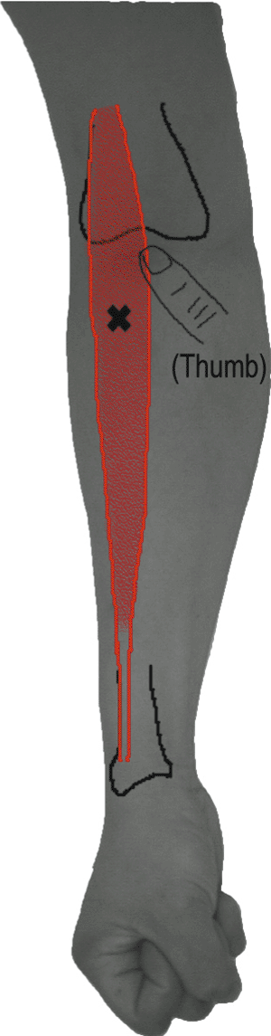
Table 8-28 Extensor Carpi Radialis (Fig. 8-28)


image
Figure 8-28 • Extensor carpi radialis
Innervation Radial nerve—posterior cord—posterior division—upper and middle trunk—C6, C7
Origin Lower third of lateral supracondylar ridge of the humerus, lateral epicondyle of the humerus, and radial collateral ligament of the elbow
Insertion Extensor carpi radialis longus: posterior aspect of base of the second metacarpal
Extensor carpi radialis brevis: posterior aspect of base of the third metacarpal
Action Extension and radial abduction of the wrist
Position Pronation of the forearm
Activation Make a fist (this avoids activation of extensor digitorum).
Needle placement Palpate the muscle adjacent to the brachioradialis on the dorsal forearm and insert one third of the distance from the lateral epicondyle to the wrist.


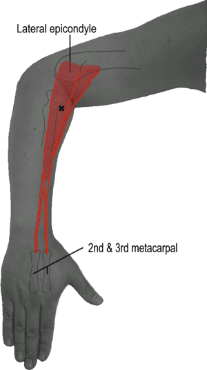
Table 8-29 Supinator (Fig. 8-29)


image
Figure 8-29 • Supinator
Innervation Posterior interosseous nerve*—radial nerve—posterior cord—posterior division—upper trunk—C6, C7
Origin Lateral epicondyle of the humerus, lateral ligament of elbow joint, annular ligament of the radius
Insertion Radial and anterior aspect of upper third of the radius
Action Supination of the forearm
Position Pronation of the forearm
Activation Supinate the forearm.
Needle placement With the forearm pronated to extend this muscle, insert 4 cm distal to the lateral epicondyle and deep (you have gone only a little too far when you hit the bone).
* Also known as deep radial nerve.


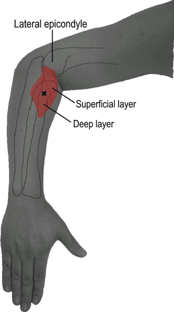
Table 8-30 Triceps Brachii (Fig. 8-30)


image
Figure 8-30 • Triceps brachii
Innervation Radial nerve—posterior cord—posterior division—middle and lower trunk—C7, C8
Origin Long head: infraglenoid tuberosity of the scapula
Lateral head: lateral and posterior aspect of the humerus
Medial head: lower posterior aspect of the humerus
Insertion Upper posterior aspect of olecranon process of the ulna
Action Extension of the elbow
Position Supine or lateral decubitus, elbow flexed
Activation Extend the elbow.
Needle placement Insert needle into each head as shown in figure. A thick fat pad overlies this muscle, making it much deeper than other muscles of the upper limb.


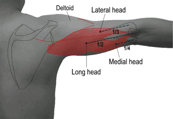
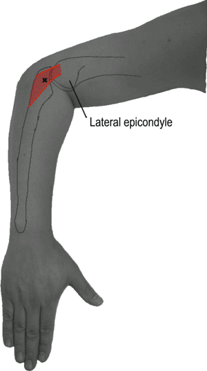
Table 8-31 Anconeus (Fig. 8-31)


image
Figure 8-31 • Anconeus
Innervation Radial nerve—posterior cord—posterior division—middle and lower trunk—C7, C8
Origin Lateral epicondyle of the humerus, posterior ligament of the elbow
Insertion Lateral aspect of the olecranon and posterior aspect of the ulna
Action Extension of the elbow
Position Pronation of the forearm with flexed elbow
Activation Extend the elbow.
Needle placement This muscle lies between the lateral epicondyle and the ulna just below the olecranon. It is the key to separating radial nerve injuries above and below the elbow.

Only gold members can continue reading. Log In or Register to continue

Stay updated, free articles. Join our Telegram channel

Sep 7, 2016 | Posted by in PHYSICAL MEDICINE & REHABILITATION | Comments Off on Pictorial Guide to Muscles and Surface Anatomy

Full access? Get Clinical Tree

Get Clinical Tree app for offline access