A thorough history and physical examination are vital to the assessment of upper extremity compressive neuropathies. This article summarizes relevant anatomy and physical examination findings associated with upper extremity compressive neuropathies.
- •
Upper extremity compressive neuropathies remain a clinical diagnosis, with scant high-level evidence to offer guidance.
- •
A thorough understanding of the anatomic course of the median, ulnar, and radial nerves are required to effectively perform a physical examination.
- •
Provocative maneuvers targeting the potential sites of compression are essential to locate the site of pathology.
- •
Electrodiagnostic testing is not without limitations, and therefore a thorough history and physical examination is essential to form the most accurate clinical picture.
Introduction
The primacy of anatomy cannot be understated with respect to the clinical diagnosis of compressive neuropathies. The examiner must assess motor and sensibility function of the nerve in question, as well as perform provocative maneuvers that may elicit neurologic symptoms. Evaluation should begin with a detailed history, as this is essential to formulate a differential diagnosis and guide physical examination.
Physical examination is fundamentally subjective; therefore, little evidence exists regarding the reliability and validity of physical examination for the upper extremity. Electrodiagnostic studies represent the best source of objective data for the diagnosis of chronic nerve compression. Electrodiagnostic testing is not without limitations, and therefore a thorough history and physical examination, with selected diagnostic testing, combine to form the most accurate clinical picture.
Median nerve
Anatomy
The medial and lateral cords of the brachial plexus, which have contributions from the sixth, seventh, and eighth cervical and the first thoracic nerve roots form the median nerve. In the upper arm, the course of the median nerve is in close proximity to the brachial artery, both of which pass along the anterior aspect of the intermuscular septum on the medial side of the arm. The median nerve and brachial artery enter the antecubital fossa medial to the biceps brachii and superficial to the brachialis muscle, then course through three successive arches as they enter the forearm. Each of these arches represents a potential site of nerve compression.
The first arch is formed by the bicipital aponeurosis (lacertus fibrosis) as it connects the biceps brachii to the flexor-pronator mass and the ulna. The median nerve is superficial to the brachialis tendon, but deep to the bicipital aponeurosis. The two heads of the pronator teres (PT) muscle form the second arch. The median nerve lies superficial to the ulnar head and deep to the humeral head. Finally, the median nerve travels between the humeroulnar and radial heads of the flexor digitorum superficialis (FDS) muscle, under the thick fibrous structure between them, known as the sublimis ridge.
In the forearm, the median nerve runs along the radial side of the flexor digitorum profundus (FDP), deep to the FDS. The anterior interosseus nerve (AIN) branches from the median nerve in the proximal half of the forearm. True to its name, the anterior interosseus nerve runs along the anterior, or volar, aspect of the interosseous membrane before terminating deep to the pronator quadratus (PQ) muscle. At approximately five cm proximal to the wrist crease, the median nerve emerges superficially between the flexor carpi radialis (FCR) tendon radially and the palmaris longus (PL) tendon ulnarly. The PL is reportedly absent in approximately 5% to 65% of the population, with wide variation across ethnic lines. The palmar cutaneous branch of the median nerve arises approximately five cm proximal to the distal wrist crease and passes outside of the carpal tunnel.
The median nerve then crosses the wrist as the most superficial of the 10 structures traversing the carpal tunnel. The transverse carpal ligament forms the roof of carpal tunnel volarly. The hook of the hamate, pisiform, and triquetrum form the ulnar wall, and the distal pole of the scaphoid and tubercle of the trapezium form the radial wall of the carpal tunnel.
Once in the hand, the thenar motor branch (or recurrent motor branch) emerges radially. The median nerve goes on to divide into radial and ulnar divisions in the plane between the flexor tendons (deep), and the palmar arch (superficially). The radial division splits to form the common digital nerve to the thumb and the proper digital nerve to the radial half of the index finger. The ulnar division splits to form the common digital nerves of the second and third web spaces.
Physical Examination
The median nerve innervates muscles involved in forearm pronation, wrist flexion, flexion of the digits, and thumb opposition and abduction ( Table 1 ). The median nerve carries sensory innervation from the radial aspect of the palm via the palmar cutaneous branch, and the volar surfaces of the thumb, index, middle fingers, and the radial half of the ring finger. Sensibility, therefore, is best tested over the thenar eminence to assess the palmar cutaneous branch and over the volar aspect of the distal index and middle fingers to assess the sensory fibers that pass through the carpal tunnel. This sensory information is essential for fine motor tasks.
| Muscle | Innervation | Action | Examination |
|---|---|---|---|
| Pronator teres | Median nerve | Forearm pronation (primary) | Resisted forearm supination, with the forearm fully pronated and the elbow extended |
| Flexor carpi radialis | Median nerve | Wrist flexion, radial deviation | Palpation of FCR with resisted wrist flexion with palpation of muscle belly |
| Palmaris longus | Median nerve | Wrist flexion (weak) | Palpation of PL tendon with resisted wrist flexion |
| Flexor digitorum Superficialis | Median nerve | PIP joint flexion | Resisted PIP joint flexion (sequential) while remaining fingers in extension, and the wrist at neutral |
| Flexor digitorum profundus (index & middle) | AIN | DIP joint flexion of the index, long, ring, and small fingers | Index DIP joint flexion while the MCP and PIP joints are held in extension |
| Flexor pollicis longus | AIN | Thumb IP joint flexion | Thumb IP joint flexion with MCP joint stabilized |
| Pronator quadratus | AIN | Forearm pronation (secondary) | Resisted forearm pronation with elbow flexed to isolate PQ from the PT |
| Abductor pollicis brevis | Median nerve | Palmar thumb abduction | Resisted abduction in the plane perpendicular to the palm with the remaining metacarpals stabilized |
| Flexor pollicis brevis (superficial head) | Median nerve | Thumb MCP joint flexion | Flexion of thumb MCP joint with IP joint held in extension to isolate from FPL |
| Opponens pollicis | Median nerve | Thumb opposition | Opposition of the volar pads of the thumb and small finger, while the examiner attempts pull the thumb away |
| Lumbricals (first and second) | Median nerve | MCP joint flexion, PIP and DIP joint extension (index and long) | Resisted extension of the PIP joint with MCP joint hyperextended (cannot isolate from interosseous muscles) |
Compressive Neuropathies of the Median Nerve
The ligament of Struthers
Approximately 1% of people have an accessory condyle or supracondylar spur approximately five cm proximal to the medial epicondyle of the humerus. The ligament of Struthers attaches this bony prominence proximally to the medial epicondyle distally. The median nerve is susceptible to compression as it passes underneath this ligament along with the brachial artery. The patient will often complain of a deep aching pain in the proximal forearm with an insidious onset, hand weakness, and numbness in the median-nerve distribution. On examination, this pain is often exacerbated with testing of the PT and FCR. Worsening of symptoms often occurs with repetitive pronation and supination. The ability to palpate this bony prominence on physical examination is variable, depending on the patient’s body habitus. Radiographs can reveal the supracondylar spur if palpation is equivocal. These patients can present with paresthesia or numbness in the median-nerve distribution as well as weakness in all muscles innervated by the median nerve, although frequently weakness of muscles innervated by the AIN is most prominent. A Tinel sign may be present proximal to the medial epicondyle. Compression of the median nerve as it passes under the bicipital aponeurosis is rare and may present similarly to compression at the ligament of Struthers.
Pronator syndrome
Pronator syndrome results from compression of the median nerve as it passes between the 2 heads of the PT. The patient often complains of aching discomfort in the forearm, weakness in the hand, and numbness in the thumb and index finger. Commonly the patient will report a history of performing forceful repetitive forearm pronation movements. On physical examination, tenderness on palpation of the PT muscle is a common finding. A Tinel sign may be present in the antecubital fossa. Testing of motor function can be difficult secondary to pain. The PT muscle receives its innervation proximal to the site of compression, and therefore might be the only muscle innervated by the median nerve spared in this syndrome. The Phalen test may be positive in 50% of patients with pronator syndrome, and therefore is unreliable in distinguishing pronator syndrome from carpal tunnel syndrome (CTS). Unlike CTS, a history of nocturnal pain and/or numbness is rare. Another provocative maneuver to test for in pronator syndrome is to apply direct pressure in the area of the PT with the patient’s forearm supinated. It is considered positive if paresthesia is reported in the median nerve distribution within one minute of compression ( Fig. 1 ).
The next possible site of compression along the course of the median nerve is the sublimis arch formed between the two heads of the FDS. Again, clinical findings are similar to those in pronator syndrome, although pain exacerbated by strong flexion of the proximal interphalangeal joints of the index, long, ring, and little fingers is suggestive of compression at the sublimis arch rather than at the PT.
Anterior interosseous nerve syndrome
AIN syndrome is an uncommon disease of unknown etiology and pathophysiology. Patients may describe vague proximal forearm pain and progressive loss in their ability to do tasks requiring fine motor control and pinch, such as handwriting. On physical examination, the patient will have weakness in the FDP to the index and middle fingers and weakness in the PQ. Deficits in the flexor pollicis longus (FPL) and index-finger FDP result in the inability to form the “OK” sign. The patient will only be able to contact the volar pads of their thumb and index fingers, rather than the tips, without flexion of the thumb interphalangeal and index distal interphalangeal joints. Sensibility is normal, as the AIN does not contain sensory fibers to the hand. Symptoms of AIN syndrome can be provoked with resisted elbow flexion, resisted forearm pronation, and resisted finger flexion ( Fig. 2 ).
Carpal tunnel syndrome
CTS results from compression of the median nerve as it travels beneath the transverse carpal ligament. The prevalence of CTS in the general population of the United States is estimated at 3.72%. CTS remains a clinical diagnosis; as there exists no gold standard test for its diagnosis. The lack of clear diagnostic standards contributes to the fact that treatment failure for CTS most commonly results from erroneous diagnosis.
Physical examination is a critical source of information in the diagnosis of CTS, although its utility as a screening tool has been questioned. Therefore, historical findings consistent with CTS are fundamental to its clinical diagnosis. Patients predominantly complain of numbness and/or paresthesia in the median-nerve distribution, rather than pain. These symptoms are typically worse at night and are improved with shaking of the hand, known as the flick sign. Patients may describe discomfort in the thenar eminence. A history of dropping objects correlates with weakness of the opponens pollicis and abductor pollicis brevis muscles. Atrophy of these thenar muscles represents advanced disease. Selected physical examination findings for the evaluation of CTS are described in Table 2 .
| Test | Procedure | Positive Result | Sensitivity/Specificity |
|---|---|---|---|
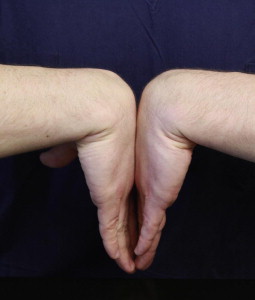
| Phalen wrist flexion test | Maximal flexion of wrists by opposing dorsal surfaces of hands ( Fig. 3 ) | Reproduction or exacerbation of paresthesia or numbness in median nerve distribution within 60 s | (0.46–0.80)/(0.51–0.91) |
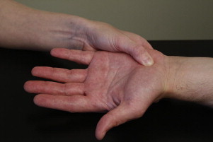
| Durkin carpal compression test | Exert direct pressure at or just proximal to the carpal tunnel ( Fig. 4 ) | Reproduction or exacerbation of paresthesia or numbness in the median nerve distribution within 60 s | (0.04–0.79)/(0.25–0.96) |
| Tinel sign | Light tapping over the median nerve at the wrist | Reproduction or exacerbation of paresthesia in the median nerve distribution | (0.28–0.73)/(0.44–0.95) |
| Static 2-point discrimination testing | Two points of various distance are applied with just enough pressure for patient to appreciate the stimulus | Inability to distinguish 2 points 5 mm apart | (0.06–0.28)/(0.98) , a |
| Semmes-Weinstein monofilament testing | Monofilaments of varying diameters are applied to volar aspect of distal index or middle finger until the filament bends | Inability to perceive a monofilament sized 2.83 or less | (0.59–0.83)/(0.59) , a |
| Ten test | A score (1–10) is reported by the patient comparing an area of abnormal light touch sensibility in the median nerve distribution to an area of similar innervation density with intact light touch sensibility | Ratio less than one between abnormal (scored 1–9) vs normal area (scored 10). Allows examiner to track changes over time | No data available |
The American Academy of Orthopaedic Surgeons (AAOS) published recommendations for diagnosing CTS based on a comprehensive review of the literature. With respect to physical examination, the level of evidence was poor because the studies are predominantly of a case-control variety, and significant variation existed in the testing maneuver protocols. The highest-level recommendation made by the AAOS group is to obtain electrodiagnostic testing if clinical evaluation is positive and surgical management is being considered. This recommendation remains controversial, as some investigators claim electrodiagnostic tests do not change the probability of diagnosing CTS in patients who are considered to have CTS based on their history and physical examination alone. In 2006, Graham used a panel of experts to assess the diagnostic utility of 57 clinical findings associated with CTS. Two historical and 4 physical examination findings had a statistically significant correlation with expert consensus diagnosis of CTS, and are referred to as the CTS-6 ( Box 1 ).
- 1.
Numbness/paresthesia predominantly in median-nerve territory
- 2.
Nocturnal numbness
- 3.
Thenar atrophy and/or weakness
- 4.
Positive Phalen test
- 5.
Loss of 2-point discrimination
- 6.
Positive Tinel sign


Median nerve
Anatomy
The medial and lateral cords of the brachial plexus, which have contributions from the sixth, seventh, and eighth cervical and the first thoracic nerve roots form the median nerve. In the upper arm, the course of the median nerve is in close proximity to the brachial artery, both of which pass along the anterior aspect of the intermuscular septum on the medial side of the arm. The median nerve and brachial artery enter the antecubital fossa medial to the biceps brachii and superficial to the brachialis muscle, then course through three successive arches as they enter the forearm. Each of these arches represents a potential site of nerve compression.
The first arch is formed by the bicipital aponeurosis (lacertus fibrosis) as it connects the biceps brachii to the flexor-pronator mass and the ulna. The median nerve is superficial to the brachialis tendon, but deep to the bicipital aponeurosis. The two heads of the pronator teres (PT) muscle form the second arch. The median nerve lies superficial to the ulnar head and deep to the humeral head. Finally, the median nerve travels between the humeroulnar and radial heads of the flexor digitorum superficialis (FDS) muscle, under the thick fibrous structure between them, known as the sublimis ridge.
In the forearm, the median nerve runs along the radial side of the flexor digitorum profundus (FDP), deep to the FDS. The anterior interosseus nerve (AIN) branches from the median nerve in the proximal half of the forearm. True to its name, the anterior interosseus nerve runs along the anterior, or volar, aspect of the interosseous membrane before terminating deep to the pronator quadratus (PQ) muscle. At approximately five cm proximal to the wrist crease, the median nerve emerges superficially between the flexor carpi radialis (FCR) tendon radially and the palmaris longus (PL) tendon ulnarly. The PL is reportedly absent in approximately 5% to 65% of the population, with wide variation across ethnic lines. The palmar cutaneous branch of the median nerve arises approximately five cm proximal to the distal wrist crease and passes outside of the carpal tunnel.
The median nerve then crosses the wrist as the most superficial of the 10 structures traversing the carpal tunnel. The transverse carpal ligament forms the roof of carpal tunnel volarly. The hook of the hamate, pisiform, and triquetrum form the ulnar wall, and the distal pole of the scaphoid and tubercle of the trapezium form the radial wall of the carpal tunnel.
Once in the hand, the thenar motor branch (or recurrent motor branch) emerges radially. The median nerve goes on to divide into radial and ulnar divisions in the plane between the flexor tendons (deep), and the palmar arch (superficially). The radial division splits to form the common digital nerve to the thumb and the proper digital nerve to the radial half of the index finger. The ulnar division splits to form the common digital nerves of the second and third web spaces.
Physical Examination
The median nerve innervates muscles involved in forearm pronation, wrist flexion, flexion of the digits, and thumb opposition and abduction ( Table 1 ). The median nerve carries sensory innervation from the radial aspect of the palm via the palmar cutaneous branch, and the volar surfaces of the thumb, index, middle fingers, and the radial half of the ring finger. Sensibility, therefore, is best tested over the thenar eminence to assess the palmar cutaneous branch and over the volar aspect of the distal index and middle fingers to assess the sensory fibers that pass through the carpal tunnel. This sensory information is essential for fine motor tasks.
| Muscle | Innervation | Action | Examination |
|---|---|---|---|
| Pronator teres | Median nerve | Forearm pronation (primary) | Resisted forearm supination, with the forearm fully pronated and the elbow extended |
| Flexor carpi radialis | Median nerve | Wrist flexion, radial deviation | Palpation of FCR with resisted wrist flexion with palpation of muscle belly |
| Palmaris longus | Median nerve | Wrist flexion (weak) | Palpation of PL tendon with resisted wrist flexion |
| Flexor digitorum Superficialis | Median nerve | PIP joint flexion | Resisted PIP joint flexion (sequential) while remaining fingers in extension, and the wrist at neutral |
| Flexor digitorum profundus (index & middle) | AIN | DIP joint flexion of the index, long, ring, and small fingers | Index DIP joint flexion while the MCP and PIP joints are held in extension |
| Flexor pollicis longus | AIN | Thumb IP joint flexion | Thumb IP joint flexion with MCP joint stabilized |
| Pronator quadratus | AIN | Forearm pronation (secondary) | Resisted forearm pronation with elbow flexed to isolate PQ from the PT |
| Abductor pollicis brevis | Median nerve | Palmar thumb abduction | Resisted abduction in the plane perpendicular to the palm with the remaining metacarpals stabilized |
| Flexor pollicis brevis (superficial head) | Median nerve | Thumb MCP joint flexion | Flexion of thumb MCP joint with IP joint held in extension to isolate from FPL |
| Opponens pollicis | Median nerve | Thumb opposition | Opposition of the volar pads of the thumb and small finger, while the examiner attempts pull the thumb away |
| Lumbricals (first and second) | Median nerve | MCP joint flexion, PIP and DIP joint extension (index and long) | Resisted extension of the PIP joint with MCP joint hyperextended (cannot isolate from interosseous muscles) |
Compressive Neuropathies of the Median Nerve
The ligament of Struthers
Approximately 1% of people have an accessory condyle or supracondylar spur approximately five cm proximal to the medial epicondyle of the humerus. The ligament of Struthers attaches this bony prominence proximally to the medial epicondyle distally. The median nerve is susceptible to compression as it passes underneath this ligament along with the brachial artery. The patient will often complain of a deep aching pain in the proximal forearm with an insidious onset, hand weakness, and numbness in the median-nerve distribution. On examination, this pain is often exacerbated with testing of the PT and FCR. Worsening of symptoms often occurs with repetitive pronation and supination. The ability to palpate this bony prominence on physical examination is variable, depending on the patient’s body habitus. Radiographs can reveal the supracondylar spur if palpation is equivocal. These patients can present with paresthesia or numbness in the median-nerve distribution as well as weakness in all muscles innervated by the median nerve, although frequently weakness of muscles innervated by the AIN is most prominent. A Tinel sign may be present proximal to the medial epicondyle. Compression of the median nerve as it passes under the bicipital aponeurosis is rare and may present similarly to compression at the ligament of Struthers.
Pronator syndrome
Pronator syndrome results from compression of the median nerve as it passes between the 2 heads of the PT. The patient often complains of aching discomfort in the forearm, weakness in the hand, and numbness in the thumb and index finger. Commonly the patient will report a history of performing forceful repetitive forearm pronation movements. On physical examination, tenderness on palpation of the PT muscle is a common finding. A Tinel sign may be present in the antecubital fossa. Testing of motor function can be difficult secondary to pain. The PT muscle receives its innervation proximal to the site of compression, and therefore might be the only muscle innervated by the median nerve spared in this syndrome. The Phalen test may be positive in 50% of patients with pronator syndrome, and therefore is unreliable in distinguishing pronator syndrome from carpal tunnel syndrome (CTS). Unlike CTS, a history of nocturnal pain and/or numbness is rare. Another provocative maneuver to test for in pronator syndrome is to apply direct pressure in the area of the PT with the patient’s forearm supinated. It is considered positive if paresthesia is reported in the median nerve distribution within one minute of compression ( Fig. 1 ).


