Osteoarthritis at the Base of the Thumb (Carpometacarpal Joint)
Min Jung Park
Jonas L. Matzon
David R. Steinberg
 A 55-year-old right-hand-dominant female laborer presents with chronic pain at the base of her thumb that worsens with pinch. |
CLINICAL PRESENTATION
Osteoarthritis of the base of the thumb is the most common arthritis in the hand that requires surgical management. Approximately 25% of women and 10% of men will ultimately develop radiographic evidence of osteoarthritis of the thumb carpometacarpal (CMC) joint. The reason for this high incidence is the unusual anatomy of the joint, which has a saddle joint architecture with little intrinsic osseous stability. This allows the thumb to move in three planes of motion (flexion-extension, abduction-adduction, and opposition) but also subjects it to high forces. Other risk factors associated with thumb CMC osteoarthritis are age, female gender, obesity, mechanical stress, hypermobility, and traumatic injuries.1
Most patients present with radial-sided hand or thumb pain that started insidiously without trauma and has been ongoing for at least a few months. The pain is exacerbated by daily activities that utilize the thumb, such as writing and turning doorknobs, and is improved with rest and pain medications. As the disease progresses, it becomes more difficult to perform tasks of daily living.
CLINICAL POINTS
Onset is usually insidious.
The condition may not have a traumatic stimulus.
More severe disease makes activities of daily living increasingly difficult.
PHYSICAL FINDINGS
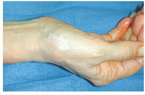
The diagnosis of CMC arthritis can usually be made by correlating the history, physical examination, and radiographs. On physical exam, inspection may reveal a prominence at the dorsoradial base of the thumb metacarpal. Palpation of this prominence will usually produce tenderness. Depending on the severity of the disease, pain may be produced by palpating the scaphotrapezial joint as well. Range of motion should be performed in all planes and compared with the contralateral side. It may be decreased secondary to pain or increased early in the disease secondary to soft tissue insufficiency. Provocative maneuvers include the CMC grind test, which involves axial thumb loading with metacarpal rotation to produce pain and trapeziometacarpal joint distraction, which stretches an inflamed capsule to reproduce symptoms. The Finkelstein maneuver (forced ulnar deviation of the wrist with flexed thumb), which is considered pathognomonic for de Quervain tenosynovitis, may also be positive in this condition.
STUDIES (LABS, X-RAYS)
Laboratory studies are generally not helpful in the diagnosis of CMC arthritis. Radiographic studies are necessary to evaluate the stage of disease. Three views of the base of the thumb are the minimum x-rays that are required. More advanced views include a basal joint stress view, which is a PA 30-degree oblique view centered on both thumbs and taken with the patient pressing the opposing thumb tips together. These x-rays can be used to stage the disease.2 In stage I, there is no joint destruction, but joint space widening secondary to synovitis may occur (Fig. 50-1). Stage II involves mild joint space narrowing with osteophytes >2 mm (Fig. 50-2). Stage III involves significant trapeziometacarpal joint space narrowing and osteophytes >2 mm (Fig. 50-3). Stage IV involves pantrapezial arthritis (trapeziometacarpal and scaphotrapezial joint involvement) (Fig. 50-4). It is important to correlate x-ray findings to patient symptoms because treatment decisions are based predominantly on symptoms. More advanced imaging such as computed tomography, magnetic resonance imaging, or ultrasound is not helpful.
NOT TO BE MISSED 
de Quervain tenosynovitis
Metacarpophalangeal (MCP), intercarpal, or radiocarpal arthritis
Compression of radial sensory nerve (Wartenberg syndrome)
Trigger thumb
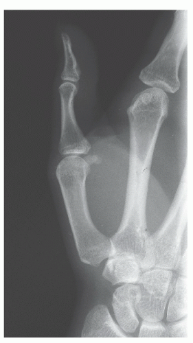 FIGURE 50-1. Stage I thumb CMC osteoarthritis, manifested by slight joint space widening. |
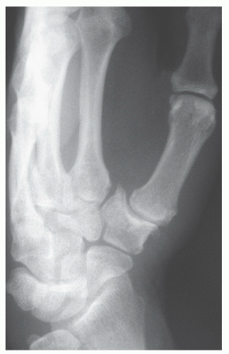 FIGURE 50-2. Stage II thumb CMC osteoarthritis, with small peripheral osteophytes and mild lateral subluxation of the joint.
Related posts:Stay updated, free articles. Join our Telegram channel
Full access? Get Clinical Tree
 Get Clinical Tree app for offline access

|
