Abstract
Polio survivors are aging and facing multiple pathologies. With age, walking becomes more difficult, partly due to locomotor deficits but also as a result of weight gain, osteoarticular degeneration, pain, cardiorespiratory problems or even post polio syndrome (PPS). These additional complications increase the risk of falls in this population where the risk of fractures is already quite high. The key joint is the knee. The muscles stabilizing this joint are often weak and patients develop compensatory gait strategies, which could be harmful to the locomotor system at medium or long term. Classically, knee recurvatum is used to lock the knee during weight bearing; however, if it exceeds 10°, the knee becomes unstable and walking is unsafe. Thus, regular medical monitoring is necessary. Orthoses play an important role in the therapeutic care of polio survivors. The aim is usually to secure the knee, preventing excessive recurvatum while respecting the patient’s own gait. Orthoses must be light and pressure-free if they are to be tolerated and therefore effective. Other joints present fewer problems and orthoses are rarely indicated just for them. The main issue lies in the prior evaluation of treatments’ impact. Some deformities may be helpful for the patients’ gait and, therefore, corrections may worsen their gait, especially if a realignment of segments is attempted. It is therefore essential to carefully pre-assess any change brought to the orthoses as well as proper indications for corrective surgery. In addition, it is essential for the patient to be monitored by a specialized team.
Résumé
La population des patients poliomyélitiques est vieillissante et polypathologique. La marche est rendue difficile par le déficit moteur induit par la maladie mais aussi la prise de poids, la dégénérescence ostéoarticulaire avec les douleurs induites, les complications cardiorespiratoires, voire le syndrome post-polio. Ces troubles de la marche sont pourvoyeurs de chutes dans une population dont le risque fracturaire est important. L’articulation clé est celle du genou. Souvent déficitaires, les muscles stabilisateurs de genou sont compensés par des stratégies de marches qui peuvent à moyen ou long terme être délétère pour l’appareil locomoteur. Le recurvatum de genou permet classiquement de verrouiller le genou lors du pas portant. S’il s’aggrave et dépasse 10°, il devient instable et ne permet plus des déplacements en sécurité. Il nécessite donc une surveillance clinique régulière. L’appareillage est le pivot de la prise en charge du patient poliomyélitique. Il s’attachera souvent à sécuriser le genou, prévenir un récurvatum excessif tout en s’inscrivant dans le schéma de marche habituel du patient. Il doit être léger sans zone d’hyper-appui pour être toléré et donc efficace. Les autres articulations sont moins problématiques et l’appareillage est rarement indiqué uniquement pour ces articulations. La difficulté réside dans l’évaluation, a priori, des retentissements du traitement proposé. Certaines déformations sont aidantes et certaines corrections envisagées, notamment celles voulant se rapprocher de la normalité anatomique, sont délétères. Toute modification d’appareillage et toute chirurgie correctrice des membres inférieurs doit être évaluée avec soins. Cette prise en charge doit être réalisée par des équipes spécialisées.
1
English version
1.1
Introduction
Gait in patients with polio sequelae can vary according to the pathological lesions, the age of onset of the initial acute attack (psychomotor development and growth) but also the age of the patient at the time of the consultation. In fact, in France, we are following a cohort of “aging” polio survivors since this pathology was eradicated at the beginning of the sixties. Nowadays, we estimate that there are 55,000 polio survivors in France . Besides, the locomotor deficit and bone deformities, we also see degenerative osteoarticular lesions of the lower limbs but also on the upper limbs promoted by the use of technical aids (crutches, canes, manual wheelchair…). This generation of patient often suffers from significant weight gain (quite detrimental to functional gait prognosis), decrease in physical activity and thus underused muscles with sometimes cardiovascular risks and/or sleep apnea syndrome or even a real diagnosis of post-polio syndrome (PPS) .
Patients develop some compensation techniques that are a real “analytical and functional paradox” leading to respecting deformations or bad postures that would seem harmful to the patient. Commonly, this becomes complicated especially when in their childhood patients had surgeries on various limbs: arthrodesis, osteotomy, muscle transfers… or a history of falls .
Another particular characteristic for the therapeutic care of these patients’ gait is to make sure that there is no sensitive nerve affection. This guarantees the lack of skin lesions in case of excessive weight bearing (on the floor or in an orthotic device) but adds to the difficulty of designing a comfortable device perfectly fitted to the patient while keeping the mechanical properties of this orthosis.
Alongside maintaining or improving the gait’s prognosis, the therapeutic strategy focuses on preventing falls and fractures. The risk of falling is related to the paralysis and the improper use of the limb. The basic principle is to respect the functional axis (for example by keeping or sometimes creating a knee recurvatum), prohibit the joint areas that promote falls such as knee flexion deformity. The risk of fractures is quite high in polio survivors. It is probably caused by the risk of falls (muscular command disorders) and bone fragility (decreased muscle tone of the affected segments, less loading on the affected limb, decreased physical activities, age…). This risk of fracture seems higher in these patients than in the general population . For poliomyelitis, it seems to affect principally the limbs rather than the spine, the upper extremity of the humerus and the lower extremity of the femur (with patella fractures). This risk of falls can be an indication to modify an orthotic device or even prescribe a new one.
1.2
Biomechanical and kinesiological issues
1.2.1
A “key” joint for the lower limb: the knee
Poliomyelitis mainly affects the knee, the triceps is more affected than the knee flexors and an isolated affection of the quadriceps is rare. It is essential to control this joint for weight bearing and gait but the knee is also the source of disorders because of the great lever arms used for resistance and the weaker lever arms for motor movements. This joint is mainly controlled by the muscles used for standing upright, the quadriceps, and a good orthopedic prognosis requires maintaining proper joint range of motion (ROM).
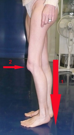
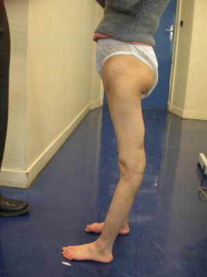
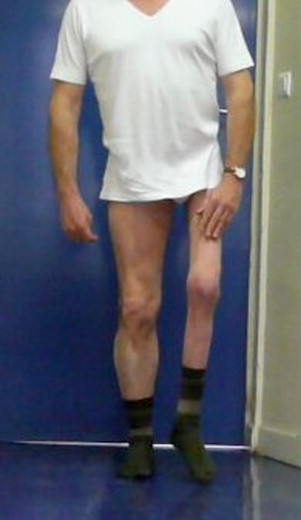
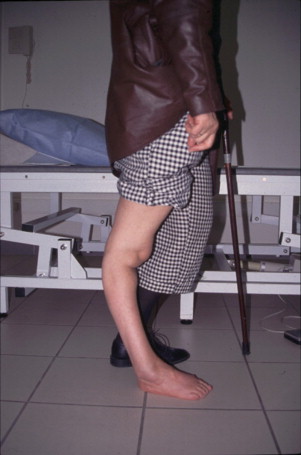
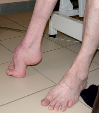
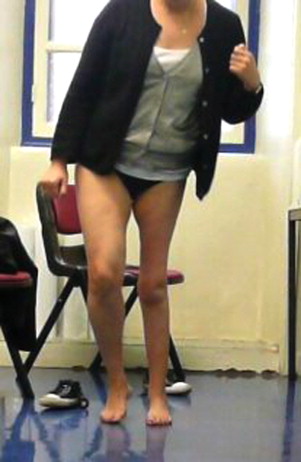
The first problem is often to comprehend how patients can lock their knee with a deficit in extensor muscles of this joint mainly weak quadriceps? In case of partial or complete paralysis of the quadriceps, loading on the lower limb is still possible if the gravity line is in front of the knee’s axis, if the capsular and tendinous structures of the knees are resistant enough and finally if the triceps or gluteus maximus muscles are adequate. Gait on a flat surface is possible because the gluteus maximus and triceps muscles compensate the quadriceps deficit only if there is a complete passive knee extension, with a slight recurvatum. The quadriceps deficit becomes a problem when going down the stairs or walking on an uneven surface. When the gluteus maximus is weak the patient must pull backwards the thigh’s axis by pushing it back with the hand or by operating a sudden extension movement of the trunk. When the triceps muscle is also weak, knee stability is bad with a risk of collapsing when the knee is in flexion. A moderate equinus deformity (10° to 20°) allows for a dynamic stability of the knee thanks to the recurvatum triggered by the tibia when the foot is flat on the floor ( Fig. 1 ). If the triceps is paralyzed, the knee stabilization is quite difficult even with adequate quadriceps. If paralysis affects several groups of muscles, knee stabilization can be reached by several compensations: complete passive extension of the knee (recurvatum); complete external rotation of the weight-bearing limb; putting the gravity line in front of the knee’s axis (Ducroquet gait pattern); stabilizing the knee in extension or in recurvatum thanks to a thigh extension triggered by the gluteus maximum muscle (associated or not to Ducroquet gait pattern); knee recurvatum triggered by foot equinus flat on the floor propelling the leg’s axis backwards ( Fig. 2 ); applying manual pressure to the thigh thus locking the thigh in extension with each step ( Fig. 3 ). These compensations are no longer possible in case of hip or knee flexion contractures, deficit of the gluteus maximus muscle and varus heel. In fact, varus heel causes excessive pressure and promotes the closing of the tibia-heel angle forward and aggravating the instability when flexing the knee.
The second problem is evaluating the long-term efficacy and impact of orthopedic changes on the knee. Knee recurvatum must be respected up to 10° and if the posterior capsular elements are resistant. Beyond 10° at 15°, knee recurvatum can get worse, become painful and create a functional shortening triggering a limp. When the posterior capsular elements are inadequate with deficient hamstrings and triceps surae, the distention of the posterior planes (progressing quickly in case of obesity) can cause a flaccid knee ( Fig. 4 ). Passive recurvatum by posterior knee structure extension, highlighted during open-chain exercise, is seen in cases of complete paralysis of the lower limbs and promoted by a fixed equinus deformity of the foot and certain compensation techniques used during gait (manual loading on the thigh). When the hamstring and triceps are functional, associated or not to an equinus deformity, and the quadriceps are deficient, the knee is pulled backward at each step, but the posterior planes are protected by the muscles and can better tolerate the constraints of this recurvatum.
1.2.2
The other joints: foot and ankle
These joints are less problematic. For the feet the deformities triggered by paralysis and imbalances generally result from the combination of several basic deformities, direct cavus foot ( pes cavus ), equinus varus foot deformity, valgus foot deformity ( pes valgus ), talipes calcaneus foot deformity, pes cavus ( Fig. 5 ), flat foot, flaccid foot and unstable foot. Upon loading the foot is often in varus position, more rarely in valgus and becomes progressively painful.
It is essential to monitor the varus deformity because it is the only way to preserve the talocrural joint (arthritis) especially if a subtalar joint and/or Chopart arthrodesis has been performed. These foot deformities are responsible for the impacts on the upper joints (knee equinus and recurvatum, knee talus and instability in flexion…) making it necessary to integrate these elements in the considering the functional disorders and their treatments.
1.2.3
Other joints: the hip
In practice this joint creates less disorders. We should note that an insufficient gluteus medius could lead to classic limps like Duchenne’s limp or swinging the shoulder on the weight-bearing side ( Fig. 6 ). An unequal length of the lower limbs (or hypoplasia) and spinal deformities (kyphosis-scoliosis) can also have an impact on the pelvis equilibrium on a sagittal and frontal plane inducing also several types of bone or joint-related limps.
1.3
Therapeutic strategies
1.3.1
Patient-acquired limps, global gait patterns and instability risks
There are several gait patterns in polio patients. The classic ones that are reported are shoulder swing limp pattern, Duchenne’s gait pattern, locked knee, walking with the hand on the thigh to lock the knee, isolated knee recurvatum, 4-step and pendulum gait patterns… All these types of gait patterns vary according to individual cases and intensity of the impairments, the use or not of technical aids (canes, orthoses…), patient’s morphology and his or her cardiorespiratory capacities during exercise.
The joint compensation strategies and gait patterns can be harmful in the long term. The strategies used over time often enable patients to make the most of their residual capacities to acquire a standing up posture and gait, often over long periods of time. However, these coping strategies can induce degenerative lesions that can become problematic to maintain a proper gait as years go by. For example, stabilizing the hip and knee by a great lateral hip lean and internal femoral rotation allow for locking the knee in an external rotation, the risk is the onset of knee dislocation in external rotation leading to unbearable arthritis.
1.3.2
Surgical solutions
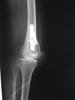
Several patients benefited from orthopedic surgery in their childhood and teenage years, mainly on the lower limbs (lengthening of the femur, osteotomy for knee recurvatum ( Fig. 7 ), femoral anteversion, arthrodesis, double even triple arthrodesis of the ankle, foot, tenotomy…). Some patients also benefited from spine surgery, mainly to control a paralytic scoliosis. For polio survivors with sequelae, some indications can be recommended. However spine surgery is limited by aging muscles and tendons, osteoarticular state (arthritic degenerescence, osteoporosis…) and by the patients’ general state (deterioration of respiratory capacities). The indications are functional ones and aim to improve gait and potential orthotic fitting, to relieve pain or avoid the aggravation of pre-existing scoliosis. The risks with these types of surgery are not negligible and experienced teams must deliver the indications. Corrective surgery can disrupt an equilibrium that was quite hard to obtain. Anatomical normality must never be a priority and the aim should be to keep the functional ability.
1.3.3
Orthotic devices
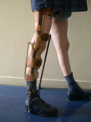
It is often during these late-setting evolutions that change in orthotics are suggested or even the prescription of a new orthotic device. Orthoses and technical aids are very useful in case of pain, joint deformities and gait disorders . A moderate knee recurvatum is thus efficient to stabilize the paralytic knee, but it tends to get worse over time especially for passive recurvatum. A deteriorating recurvatum requires a protection with an orthotic device (foot-ankle orthosis articulated around the knee with an anti-recurvatum stop) limiting the recurvatum and preventing its aggravation. The orthosis must respect some degrees of recurvatum to allow for knee stabilization and avoiding its collapse. Triceps retraction is quite common and it is important to maintain a moderate equinus foot deformity enabling the necessary recurvatum, a more important equinus deformity might be required if the knee extension is incomplete. Achilles tendon lengthening correcting the equinus foot deformity can in some cases decrease the recurvatum and make the gait more difficult. The progressive aggravation of harmful knee postures (recurvatum or valgum), muscular deficits that can sometimes be quite severe and capsular-ligament laxity can cause a flaccid knee badly tolerated in adults. In that case, the best therapeutic solution is an orthotic device: articulated knee-ankle-foot orthosis with lock ( Fig. 8 ). Wearing the latter can be an issue during the swing phase of the gait, because it is necessary to find a compromise with the uneven length of the lower limb and allow for a secure stepping phase. Knee arthrodesis must be avoided.
For the ankle and foot, the main indications are for risers or orthopedic shoes in order to gain foot stability. Sometimes an orthopedic shoe to compensate for the uneven length of the limb is recommended ( Fig. 9 ).

Bringing changes to already worn orthotics with new materials (carbon for example), for articulation, axis or loading, is not always welcomed by polio patients used to the comfort of their previous orthoses . The lack of sensitive disorders for this pathology plays an important role in this situation. Furthermore, even if it has been proved that wearing an orthosis can improve gait capacities , even in case of PPS , the indication for a new orthosis for patients who never wore one (or refused to) might not be very well accepted. It requires a lengthy discussion with the patient during which the physician will go through the potential risks without an assistive device while listening to the patient’s wishes . Acquiring technical aids is usually proposed as a first option. Often the patient has trouble accepting these aids, especially for a person who always refused to show his or her disability. Furthermore, technical aids can displace the problem to the upper limbs suffering from degenerative lesions linked to their excessive solicitation over long periods of time for gait assistance, transfers…
The role of the Physical Medicine and Rehabilitation physician will be to find medical solutions but also orthotic and surgical ones that could specifically improve the symptoms while keeping the gait abilities as long as possible according to the patients’ wishes. Designing a maladjusted orthosis can be detrimental to the patient’s gait. For example, complete orthoses of the lower limbs with a pelvic element articulated at the hip are hardly ever manufactured anymore. Their heavy weight and the disproportionate energy consumption required induced some major constraints and finally a poor tolerance.
The basic principles to abide by for prescribing orthotic devices are mainly to keep the gait pattern, greatly simplify the loading and counter-loading (no ischiatic or patellar loading if not absolutely necessary), respect the joint axis of the patient’s limbs, make sure the orthosis is as lightweight as possible, look for optimal comfort and aim for a better design in order to move away from the classic orthopedic picture, acute reminder of the “disability” era. The indication for a manual or electric wheelchair seems sometimes unavoidable, mainly for daily management of fatigue, pain (especially in the upper limbs) or weakness of the lower limbs while remembering that muscular deconditioning is one of the worst enemy for keeping muscular tonicity and autonomy . The patient’s life project should also be an intricate part of the prescription thinking process, mainly for family life, keeping up with a specific work or sport.
1.4
Conclusion
Prescribing gait orthotics for polio patients is a common problem with multiple constraints for a very heterogeneous population. In France, it is mostly designed for adult patients, often over the age of 50 presenting degenerative complications. While keeping up with global body equilibrium, the main areas of interest are the knee and mainly the loading phase of the gait cycle. Orthotic devices and their evolutions are part of the therapeutic tools for patients with PPS. In consequences, specialized teams must conduct evaluation and therapeutic care.
Related posts:
 Les attentes des patients atteints de poliomyélite antérieure aiguë
Sensitivity to change of the Quebec Back Pain Disability Scale and the Dallas Pain Questionnaire
Post-polio syndrome: Pathophysiological hypotheses, diagnosis criteria, medication therapeutics
Les attentes des patients atteints de poliomyélite antérieure aiguë
Sensitivity to change of the Quebec Back Pain Disability Scale and the Dallas Pain Questionnaire
Post-polio syndrome: Pathophysiological hypotheses, diagnosis criteria, medication therapeutics
 The importance of fear, beliefs, catastrophizing and kinesiophobia in chronic low back pain rehabilitation
The psychological aspects of polio survivors through their life experience
The importance of fear, beliefs, catastrophizing and kinesiophobia in chronic low back pain rehabilitation
The psychological aspects of polio survivors through their life experience
 Aging and sequelae of poliomyelitis
Stay updated, free articles. Join our Telegram channel

Full access? Get Clinical Tree


