Operative fixation of the scapula is associated with good outcomes. Techniques have been developed to facilitate surgical exposure of the osseous anatomy so that stability can be achieved. Although the familiar deltopectoral approach can be used for anterior glenoid fractures, the more common exposure is a posterior approach for fractures involving the neck and body of the scapula. The posterior approach has been nuanced to match needs related to fracture pattern and timing of surgery. Reducing the fragments and stabilizing them can be challenging but, a satisfactory reduction and stable fixation can be achieved, which allows immediate motion and rehabilitation.
Key points
- •
The underlying principle in the treatment of scapula fractures is that deformity and dysfunction are related as with all other fractures; therefore, restoring length, alignment, and rotation to the scapula improves outcomes.
- •
An accurate diagnostic approach to scapula fractures requires interpretation of good shoulder radiographs and a three-dimensional (3D) computed tomography scan to measure medialization at the lateral border, glenopolar angle, and scapular angulation.
- •
Extended approaches to the scapula offer excellent visualization posteriorly and should be used for complex fracture patterns or when surgery is delayed for longer than 10 days. Otherwise, more limited muscle-sparing approaches can be used that may expedite rehabilitation.
- •
There are many techniques for reduction of the lateral border, and these are paramount because it is this maneuver that is pivotal in accomplishing the surgical goals. The lateral pillar must be restored in neck and body fractures and, subsequently, rotational corrections at the medial border can be effected.
- •
The fixation montage should restore stability to the ring around the border of the scapula, from the lateral border to the spine, vertebral border, and inferior angle, so that the patient can rehabilitate effectively with immediate range of motion.
Introduction
The management of scapula fractures has evolved over recent years as new techniques and approaches have been promulgated. Often the result of a violent, high-energy mechanism, these injuries were often either missed or neglected in favor of managing higher acuity disorders. There is a 10% to 15% mortality in patients with scapula fractures, most commonly from a concomitant cranial injury or pulmonary compromise and sepsis.
A systematic review by Zlowodzki and colleagues found that, although 80% of intra-articular glenoid fractures are treated operatively, 99% of isolated body fractures received conservative therapy, at least in the first few years of this millennium. However, the field is changing rapidly, given that new reports and clinical studies published in the peer-reviewed literature have called into question the dogma that all scapula fractures do well without operative management.
Nordqvist and Petersson published the long-term results of nonoperatively treated scapula fractures and showed that 50% of patients with residual scapula deformity had shoulder symptoms. Cole and colleagues also recently published the outcomes of presurgical and postsurgical correction of extra-articular malunions of the scapula, showing a significant increase in strength and range of the shoulder, as well as an average 29-point improvement in the DASH (disabilities of the arm, shoulder, and hand) score at a mean follow-up of 3 years. As evidence mounts and the techniques improve, surgeons are becoming familiar with safe approaches, and accepting the concept of restoration of proper anatomy to achieve proper function.
Anatomy and biomechanics
An understanding of anatomy and biomechanics of the scapula is paramount for undertaking surgery of scapula fractures. Detailed descriptions can be found in an article by Cole in the January 2002 Orthopedic Clinics of North America . In brief, the scapula is a triangular bone that, in its midsection between the scapula borders, is wafer thin. There are 18 muscles that cross, originate, or insert on its borders, causing thickenings of the cortical bone that can provide purchase for screw fixation. In addition, there exists a rich vascular network, including the circumflex scapular artery along the lateral border, and a robust muscular envelope providing a salubrious biology for fracture healing; thus, nonunions are rare. The suprascapular nerve is also important to identify and protect in a posterior approach, because it traverses the spinoglenoid notch from the base of the supraspinatus fossa where it emanates from the suprascapular notch to innervate the infraspinatus ( Fig. 1 ). Traction on this nerve can cause rotator cuff weakness and should be avoided during the operation. This requirement makes it difficult to visualize the glenoid without either a tenotomy of the posterior rotator cuff or working between the interval of the teres minor and infraspinatus muscles.
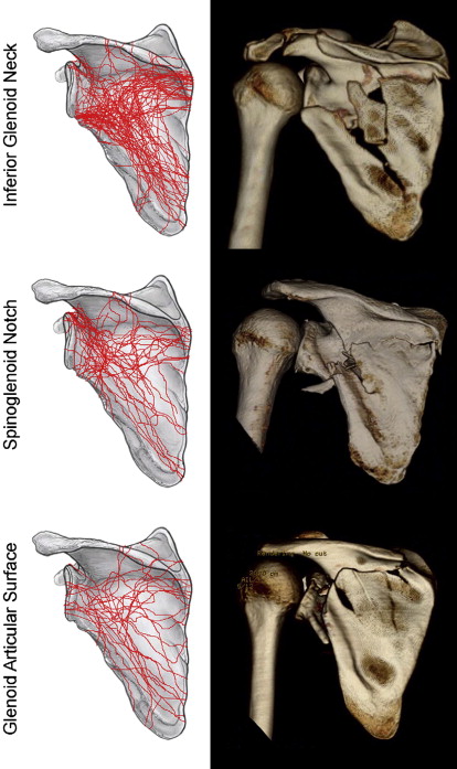
Despite the various ways the scapula can fracture, evidence suggests that most of these injuries can be codified into 3 main types. Using 3D computed tomography (CT) mapping techniques, Armitage and colleagues found in a series of operative scapula fractures that 68% of fractures involved the inferior glenoid, 17% had an intra-articular component, and 22% entered the spinoglenoid notch. In addition, of the fractures involving the inferior glenoid, most had their exit points on the superomedial border at the base of the scapula spine, and 44% had an exit at the inferomedial one-third of the vertebral border. This knowledge allows surgical planning and understanding the nature of the fracture ( Fig. 2 ).
Restoring the anatomy of the scapula facilitates a return of its biomechanical function. At its most basic level, the scapula serves to maintain the arm’s position in space. Several investigators have likened the relationship of the glenoid to the humeral head to that of a seal balancing a ball on its nose: the muscles must constantly fire and adjust to accomplish this precise feat. Furthermore, it has recently been postulated that maintaining the rotator cuff muscles at a normal tension relative to the Blick curve optimizes dynamic stability of the glenohumeral joint. Thus, by correcting the anatomy, the surgeon improves a patient’s biomechanical function.
Indications and preoperative planning
The indications used for surgery at our institution have previously been published. These indications include the following:
- 1.
Intra-articular step-off or gap greater than or equal to 4 mm
- 2.
Lateral border offset (medialization) greater than 20 mm on anteroposterior view
- 3.
Angular deformity greater than or equal to 45° seen on the scapular Y view
- 4.
Lateral border offset greater than 15 mm and angular deformity greater than 30°
- 5.
Glenopolar angle less than or equal to 22°
- 6.
Displaced double lesions of the superior shoulder suspensory complex
- a.
Both clavicle and scapula displaced greater than or equal to 10 mm
- b.
Complete acromioclavicular dislocation and scapula fracture displaced greater than or equal to 10 mm
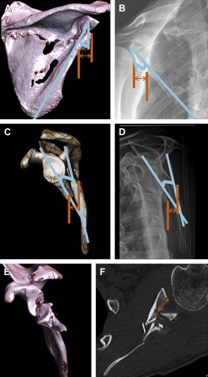
- a.
Fig. 3 shows the measurement technique to determine such indications.
Because of the nature of the mechanism of injury leading to scapula fractures, patients require a thorough preoperative evaluation, often with a multidisciplinary approach because of the polytraumatized nature of most of these patients. It is imperative that the associated life-threatening injuries receive priority, and these commonly include hemopneumothorax, cranial and spinal, as well as ipsilateral extremity injuries. The associated injury rate in displaced operative scapula series is as high as 90%. In addition, a comprehensive secondary survey performed and documented by the orthopedic team is essential to rule out other injuries that may require orthopedic care. Of particular concern is the strong incidence of brachial plexus palsy, approximately 12.5% associated with scapular fractures, and about a 50% ipsilateral extremity injury rate. A neurologic examination can be challenging in these patients because of intubation in many cases, pain, and ipsilateral fractures, but nevertheless should be completed with vigilance, because the neurologic status can adversely affect outcome.
The preoperative evaluation also involves a soft tissue assessment of the integument, because many of the fractures occur from a blow to the superior shoulder, which can be associated with abrasions. We prefer to defer surgery until abrasions have completely epithelialized to reduce risks of infection, and, to date, we have not had an infection for this surgery, an astounding statistic that happens to mimic the literature on operative series. Abrasions may take 3 to 4 weeks to resolve, which is enough time for the scapula to form significant callus. Results of surgery in 22 such delayed patients having open reduction with internal fixation were associated with good outcomes as assessed with DASH, as well as range of motion and strength. A daily Hibicleanse scrub is used to reduce the bacterial load over the compromised area. Neither topical nor oral antibiotics are necessary during this phase.
Preoperative planning also must include an assessment of the fracture radiographically and preferably with CT 3D reconstructions both to assess deformity and plan surgical approaches. It is useful to obtain an anteroposterior view of the opposite shoulder to measure the normal glenopolar angle, to compare the restored fractured side with regard to this important alignment variable, which is discussed later.
Surgical Approaches
The chosen surgical approach depends on fracture location, complexity, and chronicity. Less invasive approaches are preferred when feasible, but are technically more demanding. There is an unstudied belief that patients rehabilitate faster and have less pain with more limited dissection. The scapula heals quickly because of the rich local blood supply, but surgery is often delayed because of the treatment of other critical injuries or lost time in transferring the patient to an expert. Therefore, the extensile approach is utilitarian and often used for such situations, because complete exposure of the scapula may be necessary in such circumstances to mobilize callus.
Posterior Approaches
Although the posterior approach has been considered the workhorse for any fracture involving the scapula body or neck, there have recently been several nuances to the posterior approach that have been described. A posterior approach should be used except in the case of anterior glenoid fractures, or superior glenoid fractures involving the coracoid, or isolated process fractures.
Variations on posterior approaches include (1) extensile Judet approach, (2) modified Judet approach, and (3) the minimally invasive direct approach. The senior author (PAC) is now more frequently using an open Judet approach, which spares the deltoid from takedown allowing full access, and has also developed the minimally invasive technique for fractures that are acute and simple. In all cases, the setup and equipment are the same.
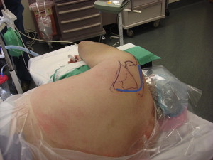
The patient is positioned on a bean bag in the lateral decubitus position. A triangular BoneFoam positioner (BoneFoam Inc, Plymouth, Minnesota) is placed on a hand table to allow the ipsilateral extremity to be prepped and draped free and manipulated as necessary during the case ( Fig. 4 ), which facilitates manipulation during the operation, as well as allowing the patient to flop slightly forward, which is instrumental in improving the surgeon’s visualization and access to the scapula. The forequarter is prepped from midchest to midback, and from the neck to the flank. Should the patient also possess an ipsilateral clavicle fracture, this can be addressed without reprepping and draping by simply maneuvering the arm out of the so-called floppy-forward position, and the surgeon taking a cephalad and anterior position to execute this part of the surgery.
The following implants and instrumentation are useful for surgical exposure and reduction of the fractures and represent the preference of the senior author ( Fig. 5 ):
- •
2.0-mm, 2.4-mm, and 2.7-mm plates for most cases
- ○
Although locking implants may be useful, they are not necessary. They help to decrease the need for longer plates and minimize incision sizes.
- ○
Both 2.7-mm reconstruction plates for the spine and vertebral border and angle of the scapula as well as 2.7-mm dynamic compression (dc) plates for the lateral border.
- ○
- •
Small external-fixator set with 4 mm Schanz pins
Shoulder hook
- •
Cobb elevator
- •
Large and medium Hohmann retractors and Deaver retractors
- •
Lamina spreaders of various sizes; pituitaries of various sizes; #2 braided nonabsorbable sutures, as well as #1, 0, 2-0 absorbable braided sutures, and an absorbable monofilament for the skin, and a 3.2-mm to 6.4-mm (1/8-inch to 1/4-inch) self-suction drain
Related posts:
 Results and Outcomes of Unicompartmental Knee Arthroplasty
Technical Pitfalls of Shoulder Hemiarthroplasty for Fracture Management
Posterior Elbow Wounds
Management of Radial Nerve Palsy Following Fractures of the Humerus
Technical Pitfalls of Shoulder Hemiarthroplasty for Fracture Management
Reverse Shoulder Arthroplasty
Results and Outcomes of Unicompartmental Knee Arthroplasty
Technical Pitfalls of Shoulder Hemiarthroplasty for Fracture Management
Posterior Elbow Wounds
Management of Radial Nerve Palsy Following Fractures of the Humerus
Technical Pitfalls of Shoulder Hemiarthroplasty for Fracture Management
Reverse Shoulder Arthroplasty
Stay updated, free articles. Join our Telegram channel

Full access? Get Clinical Tree


