Operating Room Preparation and External Fixation Equipment
Paul S. Cooper
Introduction
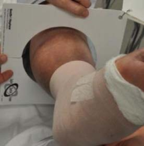
Perioperative planning can significantly improve the efficiency of external fixator application in the operating room. Template ring sizing can reduce inventory on the back operating table during setup and the surgeon should have a preliminary idea of the type and design of external fixation needed for the procedure (Figure 5.1). In many of the foot and ankle cases, the circular external fixators can be pre-built and sterilized, expediting the operating room time for application (Figure 5.2A and B).
Operating Room Preparation for Circular External Fixation of the Foot and Ankle
The operating table should be positioned in the room so that there is adequate sterile space at the distal end for surgeon movement during multiplane wire insertion. The patient is positioned on a standard operating room table if mini C-arm fluoroscopy is utilized. Alternatively, more proximal cases involving the tibia, for example, corticotomy with bone transport, are best performed on a fluoroscopy operating table. Mini fluoroscopy is positioned on the ipsilateral surgical side, while a conventional image intensifier C-arm fluoroscope is positioned on the contralateral side (Figure 5.3).
 Figure 5.1. Preoperative ring sizing of the lower extremity determines which rings to have available in the operating room. |
The patient is positioned with the operative lower extremity in neutral position utilizing bolsters under the ipsilateral buttocks and/or shoulder (Figure 5.3). The contralateral lower extremity is well padded over the fibular head and malleoli to prevent peroneal nerve compression and pressure sores over the lateral ankle (Figure 5.4). The use of a pneumatic sequential compression device for the contralateral lower extremity is considered on the basis of the time duration of the surgery. A thigh tourniquet is placed to allow full access to the tibia and knee joint. The patella should be draped out and needs to be visualized to access for rotation, and the foot should be located a few centimeters proximal to the end of the operating table. The leg may need to be elevated on a leg holder to facilitate lateral fluoroscopic examination during the procedure (Figure 5.4). Draping should include multiple layers to minimize transosseous wire penetration during circular external fixation application. Additional sterile towels should be available during the case for positioning of the lower extremity within the circular external fixation device.
External fixation application comprises multiple components often requiring assembly intraoperatively. An organized back table is essential for efficient building and application of the external fixation device (Figure 5.5). The back table should have the basic trays of components laid out, or to increase efficiency, a larger single tray with the most frequently used components (Figure 5.6A–C). If half-pins are planned to be utilized, a separate tray with pins, insertion tools, and pin cutters is used (Figure 5.7). A pre-built circular external fixation that can be utilized for most foot and ankle cases is placed on the back table, or alternatively, a ring tray is added for custom circular external fixation build out intraoperatively. A separate table is set up for standard instruments including power saws and burrs, osteotomes, and retractors. Extra batteries for the power instrumentation should be readily available. Maintenance of
separation between instrumentation used to address infected or contaminated tissues and those used during the circular external fixation application is critical to reduce risk of postoperative complications.
separation between instrumentation used to address infected or contaminated tissues and those used during the circular external fixation application is critical to reduce risk of postoperative complications.
Stay updated, free articles. Join our Telegram channel

Full access? Get Clinical Tree


