Open Reduction and Internal Fixation of Intra-articular Scapular Fractures
Brett D. Owens
Joanna G. Branstetter
Thomas P. Goss
DEFINITION
Intra-articular scapular fractures include fractures of the glenoid cavity, which includes the glenoid rim and the glenoid fossa. They account for 10% of scapular fractures.6 Most scapular fractures are extra-articular, and 50% involve the body and spine.
Over 90% of fractures of the glenoid cavity are insignificantly displaced and are managed nonoperatively.3
Significant displacement requires evaluation for surgical intervention to achieve the best possible outcome.
ANATOMY
The scapula is a flat triangular bone with three processes: the glenoid process, the acromial process, and the coracoid process.
The glenoid process consists of the glenoid cavity (the glenoid rim and glenoid fossa) and the glenoid neck.
The glenoid cavity provides a firm concave surface with which the convex humeral head articulates. The average depth of the articular cartilage is 5 mm.
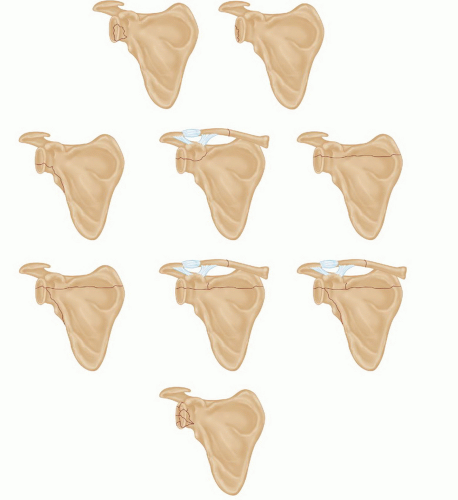
Glenoid cavity fractures are classified according to whether they involve the glenoid rim or the glenoid fossa and the direction of the fracture line (FIG 1).
PATHOGENESIS
Scapular fractures usually are the result of high-energy trauma and have a high rate (90%) of associated bony and soft tissue injuries, both local and distant.5
Fractures of the glenoid rim occur when the humeral head strikes the periphery of the glenoid cavity. They are true fractures, not avulsion injuries caused by indirect forces applied to the periarticular soft tissues by the humeral head.
Fractures of the glenoid fossa occur when the humeral head is driven into the center of the concavity. The fracture then promulgates in a number of different directions, depending on the characteristics of the humeral head force.
NATURAL HISTORY
The results of nonoperative treatment of intra-articular scapular fractures usually are good if the fracture displacement is minimal and the humeral head lies concentrically within the glenoid cavity.
Significant displacement can result in posttraumatic degenerative joint disease, glenohumeral instability, and even nonunion.2
PATIENT HISTORY AND PHYSICAL FINDINGS
In addition to the specifics of the injury, it is helpful to obtain an understanding of the functional demands on the extremity. Hand dominance, occupation, and sports participation are all relevant.
A thorough neurovascular examination must be performed.
Deficits are evaluated with angiography and electromyography, as necessary.
A thorough soft tissue examination also is warranted. Wounds may represent an open fracture and warrant exploration. Blisters or swelling may delay surgery.
IMAGING AND OTHER DIAGNOSTIC STUDIES
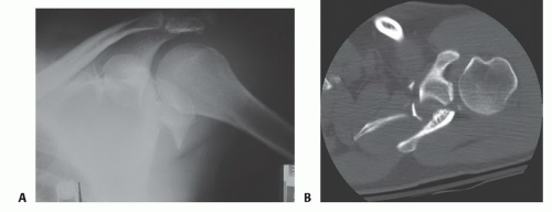
Intra-articular scapular fractures initially are evaluated with a routine scapula trauma radiographic series (a true anteroposterior view of the shoulder with the arm in neutral rotation, a true axillary view of the glenohumeral joint, and a true lateral scapular view; FIG 2A).
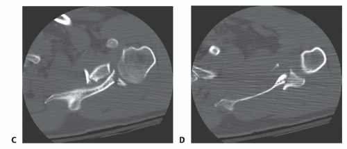
Computed tomography (CT) scans and three-dimensional studies with reconstructions can be helpful in evaluating articular congruity and fracture displacement (FIG 2B-D). In addition, the bony relationships should be evaluated for evidence of ligamentous disruption(s) or instability.
DIFFERENTIAL DIAGNOSIS
Intra-articular scapular fractures
Nonarticular scapular fractures
Scapulothoracic dissociation
Double disruptions of the superior shoulder suspensory complex, including a floating shoulder (a glenoid neck fracture with an ipsilateral middle third clavicle fracture)
NONOPERATIVE MANAGEMENT
Most (over 90%) intra-articular scapular fractures are insignificantly displaced and are managed nonoperatively.
Significantly displaced glenoid fossa and glenoid rim fractures require operative management.
SURGICAL MANAGEMENT
Surgical indications are as follows:
Rim fractures: 25% or more of the glenoid cavity anteriorly or 33% or more of the glenoid cavity posteriorly and displacement of the fragment 10 mm or more
Fossa fractures: an articular step-off of 5 mm or more, significant separation of the fracture fragments, or failure of the humeral head to lie in the center of the glenoid cavity
Preoperative Planning
Imaging studies should be reviewed before the surgery and should be available for reference in the operating room. A draped fluoroscopy unit and a competent technician should be available. An examination for instability can be performed while under anesthesia.
Positioning
Open reduction with internal fixation (ORIF) of intra-articular scapular fractures requires wide access to the entire shoulder girdle. Depending on the particular fracture, the patient is placed in either the lateral decubitus position (FIG 3A) or the beach-chair position (FIG 3B).
Care must be taken to allow adequate exposure of the entire scapula and clavicle. The shoulder girdle is prepped and draped widely, and the entire upper extremity is prepped and draped “free.”
In some cases, a staged procedure may be necessary using separate positions, sterile preparations, and exposures.10
Stay updated, free articles. Join our Telegram channel

Full access? Get Clinical Tree


