Open Reduction and Internal Fixation of Diaphyseal Forearm Fractures
Lee M. Reichel
John R. Dawson
DEFINITION
Diaphyseal forearm fractures include isolated or combined radial and ulnar fractures (“both-bone fractures”). They occur distal to the elbow joint and proximal to the wrist joint.
It is critical to evaluate the distal radioulnar joint (DRUJ) and radiocapitellar joint preoperatively, intraoperatively, and postoperatively to avoid missing Galeazzi- and Monteggia-type injuries.
Fixation techniques should be tailored to the age of the patient and the location and pattern of fracture.
Excellent functional results and union rates can be obtained when skeletal length and alignment are restored with stable internal fixation.
ANATOMY
Complete knowledge of neural, vascular, and muscular anatomy is expected. Neural anatomy is particularly important, as a nerve injury in the forearm rarely completely recovers. Nerve injuries result in disabling, temporary or permanent, motor and sensory dysfunction in the hand.
Injury
Radial, posterior interosseous (PIN), median, anterior interosseous (AIN), and ulnar nerve injuries can all occur, although their incidence is not frequent. Preoperative nerve assessment is best performed by measurement of static two-point discrimination. Acutely, motor examinations are difficult secondary to pain. If a nerve injury is suspected preoperatively, that nerve must be explored within the zone of injury. Although the majority of nerves are found to be in continuity, the surgeon should be prepared to repair the nerve either primarily or with nerve cable grafts following bony stabilization.
Unless injured preoperatively, the radial, median, and ulnar nerves are not typically encountered. If they are encountered, this should alert the surgeon that he or she might be in the wrong dissection interval.
Muscle injury may be significant following fracture. It is typically not clinically significant except for injury to the flexor pollicis longus, which may even be nonfunctioning in severe injuries. This can be difficult to differentiate preoperatively from a partial AIN injury.
Approaches to the radius (FIG 1)
Five muscles cover the radius (supinator, flexor digitorum superficialis, pronator teres, flexor pollicis longus, pronator quadratus). When the soft tissue injury is significant, muscle size, fiber orientation, and tendinous insertions (particularly the pronator teres) help orient the surgeon. The supinator muscle is especially important to identify in both volar and posterior approaches to the radius to avoid injury to the PIN. Its fibers are obliquely oriented to the longitudinally oriented flexor and extensor muscles.
During the volar, or anterior, approach, the lateral antebrachial cutaneous nerve, superficial radial sensory nerve, AIN, and PIN are usually encountered. The lateral antebrachial cutaneous nerve is sometimes encountered during blunt scissor dissection through the subcutaneous fat following the skin incision. Proximally, the superficial radial nerve lies deep to the brachioradialis. One must avoid placing self-retaining retractors on it.
The radial artery is encountered in every anterior approach to the radius. It is found deep to the brachioradialis in the proximal one-third of the forearm and visualized just beneath the forearm fascia exiting near the divergence of the brachioradialis and flexor carpi radialis muscle bellies in the midforearm. In very proximal volar approaches, near the bicipital tuberosity, crossing veins and the recurrent radial artery can be visualized.
Superficial veins of the volar and dorsal forearm can be large and contribute to significant bleeding. Formal suture ligation may be needed for large veins.
During the dorsal, or posterior, approach to the radius, the PIN and possibly the superficial radial sensory nerve may be encountered.
Approaches to the ulna (see FIG 1)
The dorsal ulnar cutaneous nerve is most commonly visualized passing in a volar to dorsal direction through the subcutaneous tissue, distal to the ulnar styloid. However, rarely, variations do exist where this nerve crosses the ulna more proximally. Therefore, blunt dissection through the subcutaneous fat in the distal one-third of the forearm is safest for preventing inadvertent nerve injury.
The entire ulna is a subcutaneous bone, and subperiosteal dissection provides extensile exposure. Flexor carpi ulnaris and flexor carpi radialis border the volar and dorsal sides of the ulna. These muscles converge in the middle third of the ulna, requiring only shallow intramuscular dissection to expose the ulnar shaft.
Fixation
Both the AIN and PIN lay millimeters away from the radius in anterior and posterior approaches. Reduction clamps inadvertently
placed around them when affixing a plate to the bone or reducing fracture fragments can damage them. Additionally, avoid using monopolar cautery on the ulnar aspect of the radius. When bleeding is encountered from the anterior interosseous vessels, they must be dissected away from the nerve prior to obtaining hemostasis to avoid nerve injury. Bleeding is stopped with bipolar cautery or small vascular clips.
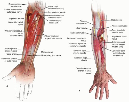
FIG 1 • Muscular and neurovascular anatomy of the forearm. A. During a volar approach to the forearm, the radial artery, superficial radial nerve, anterior interosseus neurovascular structures, and posterior interosseous nerve may all be encountered. Detailed knowledge of their location and ability to visually identify these structures are critical to avoiding injury when their anatomic location is disrupted by injury. B. Dorsal approaches must demand identification of the posterior interosseious nerve proximally and superficial radIal nerve branches distally. During distal third ulnar approaches, the dorsal cutaneous branch of the ulnar nerve may be encountered notably when anatomy is aberrant.
Osteology
The radius has a complex osteology with both a radial and sagittal bow. The radial bow has an arc of approximately 10 degrees and lies in the coronal midshaft, whereas the sagittal bow has an approximately 5 degree arc and lies in the proximal third of the radius.9 Contouring of anteriorly placed plates on the proximal radius accommodates the sagittal bow. Anatomic plates are available to accommodate the radial bow.
The ulna is generally flat in the sagittal plane and curved in the coronal plane (with the exception of the proximal ulna which in some patients has a slight apex posterior curvature at the olecranon).8 In the middle and distal thirds of the forearm, plate fixation can be placed anteriorly or posteriorly to avoid symptomatic hardware. In proximal ulnar shaft fractures, plate placement along the subcutaneous border, although possibly more symptomatic, obviates the need for plate contouring to the ulnar coronal bow. This placement also helps resist the forces generated during elbow flexion and extension from the long lever arm of the forearm.
PATHOGENESIS
Direct trauma (guarding face against direct blow, gunshot wound)
Indirect trauma (motor vehicle collision, falls)
The incidence of associated injuries in patients presenting to a trauma center with a both-bone forearm fracture is significant. In one series of 87 patients presenting to a regional
trauma center, 40% had multiple injuries (25% with closed head injury, 26% associated major injuries in the same extremity).3
NATURAL HISTORY
Closed treatment of radius or both-bone forearm fractures generally yields unacceptable results.1
Plate fixation using 3.5-mm compression plates of radial and ulnar fractures is the standard of care yielding good or excellent functional results and union rates greater than 95%.3
Restoration of forearm rotation depends on obtaining proper skeletal length and axial and rotational alignment.11
PATIENT HISTORY AND PHYSICAL FINDINGS
Evaluate for life-threatening injuries first.
When there is obvious injury to the forearm, it should be examined last so that satisfaction of search does not result in missed injuries.
Examination begins at the neck and shoulder girdle away from the injured area. In an awake, cooperative patient, palpation of each bony structure will typically reveal injury for which imaging should be obtained. In an uncooperative or intubated patient, a very low threshold for obtaining imaging is needed.
It is particularly important to palpate the radial head, collateral ligaments of the elbow, distal radius and ulna, and triangular fibrocartilage complex to avoid missing soft tissue, Monteggia, or Galeazzi injuries. If a ligamentous or tendinous injury is suspected in the setting of a stable joint, a magnetic resonance imaging (MRI) scan is ordered to make the diagnosis and allow for early repair if indicated.
Usually, obvious gross deformity is present when both the radius and ulna are fractured, but isolated radius or ulna fractures are easily missed especially in a polytrauma, intubated, or noncommunicative patient.
It is critical that the forearm compartments be visualized in their entirety and palpated to assess for compartment syndrome. All splints and dressings must be removed so the skin can be examined circumferentially. The signs and symptoms of compartment syndrome should be checked and documented even when they are “negative.”
The neurovascular examination at a minimum should include an assessment of radial and ulnar pulses and a detailed documented examination of the sensorimotor function of the median, radial, and ulnar nerves. Preoperative AIN function should be documented as well.
IMAGING AND OTHER DIAGNOSTIC STUDIES
Anteroposterior and lateral radiographs of the forearm, wrist, and elbow generally suffice.
Careful scrutiny of the DRUJ and radiocapitellar joint alignment are performed on wrist and elbow radiographs.
In comminuted fractures, contralateral imaging of the uninjured forearm and wrist is helpful to determine the patient’s native bony alignment and ulnar variance.
DIFFERENTIAL DIAGNOSIS
Radial shaft fracture with DRUJ injury (Galeazzi fracture)
Ulnar fracture with radiocapitellar dislocation (Monteggia fracture)
Compartment syndrome
NONOPERATIVE MANAGEMENT
Nonoperative care is reserved for middle or distal third isolated ulnar fractures with no associated injury to the proximal radioulnar joint (PRUJ) or DRUJ. Proximal fractures are rarely treated nonoperatively.
Generally, greater than 50% of bony overlap and less than 15 degrees of angulation are appropriate for nonoperative management.
Distal fractures can be maintained in a fracture brace or short-arm cast. Midshaft fractures can be immobilized in “Munster cast” as described earlier or in a fracture brace.
The duration of immobilization is until pain subsides and the patient can tolerate mobilization. Weight bearing through the extremity is avoided until there is clinical and radiographic evidence of fracture union. Early mobilization may lead to more rapid union.2
Rarely, stable isolated nondisplaced radius shaft fractures can be treated in a cast or functional brace that allows elbow flexion and extension but no forearm rotation.
Radiographic union can be expected between 8 and 10 weeks.
SURGICAL MANAGEMENT
The two primary goals of treatment are to obtain union and restore function. The primary surgical aim is the stable restoration of length, angular alignment, and rotational alignment.
Approach
Separate approaches are needed for the radius and ulna to minimize the risk of synostosis.
Radius fixation is performed through an anterior or a posterior approach. Anterior fixation minimizes but does not eliminate the possibility for symptomatic hardware. Anterior fixation is straightforward for middle-third and distal-third radius fractures but is more difficult in proximal-third fractures. The posterior approach has traditionally been recommended for the middle-third radius fracture but is rarely used. The posterior approach is most helpful during proximal radius exposure but care needs to be taken to protect the PIN.
The entire ulna can be exposed through a subcutaneous approach. Plate fixation can be on the subcutaneous surface, anterior surface, or dorsal surface.
Internal fixation
The order of operation in both-bone forearm fractures depends on the degree of comminution of each bone. Typically, the less comminuted bone is fixed first so as to have the most precise restoration of length.
Radius fractures are stabilized with the arm extended, whereas the ulna is typically stabilized with elbow flexed 90 degrees. Therefore, if indicated, radius fixation first allows a stable forearm during elbow flexion for ulnar fixation.
3.5-mm compression plates with six cortices of fixation on either side of the fracture are the standard of care. Anatomic and straight plates are available with locking and nonlocking screw options. Comminuted fractures may require bridge plating. Anatomic plates are very helpful for restoring the radial bow.
In osteoporotic fractures, the use of locking screws is indicated.
Locked plates are also indicated when bridging a defect or when one segment is very short and six cortices of fixation cannot be achieved.
Care must be taken to ensure plates and screws placed on the both the distal ulna and the proximal radius does not impinge in each respective radioulnar joint. Locked unicortical screws must sometimes be used to avoid screw tip prominence in the joint. Live intraoperative fluoroscopic examination is used to assessing screw placement near the DRUJ or PRUJ. Additionally, it is critical to pronosupinate the forearm to ensure there is no plate impingement in these joints.
Bone grafting
Closure
The tourniquet must always be taken down and meticulous hemostasis obtained.
Fascia is left open and only skin and subcutaneous tissues are approximated.
When there is significant swelling that results in a tight closure, skin should be left open, and a delayed primary closure is usually obtainable after 72 hours.
Soft well-padded dressings with no tight circumferential wrapping minimize the risk of postoperative compartment syndrome.
Special circumstances
If the patient presents with signs and symptoms of compartment syndrome, they should be taken STAT to the operating room for decompressive fasciotomy of the forearm and carpal tunnel at a minimum. Typically, a single volar incision fasciotomy with release of superficial and deep fascial structures will suffice in decompressing the forearm compartments. Mobile wad and dorsal extensor compartments and hand compartments need to be critically evaluated following volar fasciotomy. They should be released if any suspicion exists for compartment syndrome of these compartments.Related posts:
 Open Reduction and Internal Fixation of Displaced Lateral Condyle Fractures of the Humerus
Open Reduction and Internal Fixation of Capitellum and Capitellar-Trochlear Shear Fractures
Open Reduction and Internal Fixation of Ulnar Styloid, Head, and Metadiaphyseal Fractures
Fragment-Specific Fixation of Distal Radius Fractures
Vascularized Bone Grafting and Capitate Shortening Osteotomy for Treatment of Kienböck Disease
Operative Treatment of Metacarpal Fractures
Open Reduction and Internal Fixation of Displaced Lateral Condyle Fractures of the Humerus
Open Reduction and Internal Fixation of Capitellum and Capitellar-Trochlear Shear Fractures
Open Reduction and Internal Fixation of Ulnar Styloid, Head, and Metadiaphyseal Fractures
Fragment-Specific Fixation of Distal Radius Fractures
Vascularized Bone Grafting and Capitate Shortening Osteotomy for Treatment of Kienböck Disease
Operative Treatment of Metacarpal Fractures

Stay updated, free articles. Join our Telegram channel

Full access? Get Clinical Tree


