Neurovascular Syndromes of the Extremities


Neurovascular Syndromes of the Extremities


As I define them, the regional neurovascular syndromes of the extremities are rare disorders. That is because I insist that these diagnoses not be made by exclusion. For me to have confidence in these diagnoses, there must be specific, even quantifiable, abnormalities demonstrable by reliable objective testing. In a few instances, generally instances of severe involvement, there may be specific findings on physical examination. However, short of these criteria, I am unwilling to use any of the neurovascular diagnostic labels.

There are other observers, authors, and clinicians who take issue with my stridency. They argue that there must be forme fruste of all these conditions, that is, patients who are afflicted with an illness but not with such severity to manifest anything more than symptoms. Of course, this argument is valid. However, it is not defensible at the bedside for the clinician, in the field for the epidemiologist, and certainly not in the halls of policy. Many regional musculoskeletal disorders of the extremities are far more prevalent, by orders of magnitude, than the neurovascular syndromes. As discussed in the previous chapters, most of the regional disorders of the extremities are diagnosed by exclusion. And many of these conditions present with symptoms that overlap those of the neurovascular syndromes. The upshot is that the diagnosis of forme fruste neurovascular syndrome is highly likely to be wrong; it is far more likely to represent mislabeling of another regional disorder. This caveat may hold even in those instances in which objective testing is available to diagnose a neurovascular syndrome. I will illustrate this further in the discussion of carpal tunnel syndrome (CTS) later in the chapter when I demonstrate how the prior probability that your patient has CTS influences the utility of any diagnostic testing.

I am so strident about the need to restrict labeling of neurovascular syndromes to individuals with specific symptoms and objective abnormalities that I will shortly argue for discarding a time-honored label, primary Raynaud’s, because it denotes nothing more than a normal variant.

Of course, my strident diagnostic posture means I will miss out on the lucky happenstance of appropriately labeling the patient whose illness does represent forme fruste of a discrete neurovascular syndrome. Seldom does this compromise the patient. In most instances of very early or mild disease, all that is called for are the conservative measures that apply to the regional disorders and expectant observation. As a diagnostician, expectant observation is my burden and the reason for my anxiety. There is no reason to confound my patient’s coping by sharing degrees of uncertainty that are so low on statistical grounds. Rarely will a regional
disorder prove, over time, to have been the early presentation of a neurovascular syndrome or, for that matter, a systemic rheumatic disease.


ENTRAPMENT NEUROPATHIES

The major peripheral nerves of the upper and lower extremities are at risk for compression and consequent physiologic compromise at particular sites in their course. Most such entrapment neuropathies are “idiopathic”; they occur without a discrete precipitant and are not associated with any underlying disease process. However, the nerves are at risk at these same sites and other sites for traumatic disruption in the setting of major tissue damage or major blunt trauma. There are surgical treatises on this topic. The current discussion is designed to provide a clinically relevant foundation of information relevant to regional disorders and their differential diagnosis.

Rheumatoid synovitis can also impinge on the peripheral nerves as they traverse foci of inflammation, generally at the same points of susceptibility as seen in idiopathic entrapments.1 Other systemic diseases either wreak havoc at specific sites or, in the case of infection or neoplasm, in the presence of pathologic tissue. We will mention some of these as we discuss particular entrapments.

There are features of the clinical presentation common to all entrapment neuropathies:


  • Dysesthesias are characteristically localized to the sensory distribution of the nerve.


  • Discomfort and paresthesias are more prominent at rest than with usage.


  • There is greater susceptibility of sensory fibers than motor fibers to the insult.


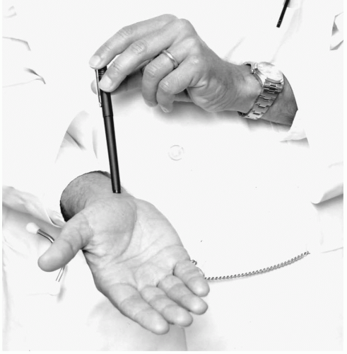
  • Tinel’s sign is frequently present. This means that percussion of the nerve at the site of entrapment elicits dysesthesias in the sensory distribution of the nerve.


  • Electrodiagnostic studies provide the gold standard for all entrapment neuropathies.

The reliability of all these features, including electrodiagnostic findings, varies from site to site and as a function of the pathogenesis. The utility varies as a function of the likelihood (prior probability) that a given patient is actually afflicted (vide infra).

The pathogenesis of the idiopathic entrapment neuropathies is under intensive study. Normally, peripheral nerves withstand the impressive microvascular changes that can be induced with relatively low pressures. However, with prolonged or increased pressure and deformation, the zone between the compressed and the free nerve suffers a permeability change leading to interfascicular edema, a steeper pressure gradient, and more pronounced impedance to conductivity. Large-diameter fibers are more susceptible to compression than smaller fibers. Nerves that have suffered from other processes, such as the microcirculatory disease of diabetes, are particularly at risk for prolonged and even permanent damage.


Thoracic Outlet Syndrome

Thoracic outlet syndrome is a concept with a colorful history that engenders controversy to this day.2 Near the turn of the 20th century it was suggested that the neurovascular structures that comprise the brachial plexus and associated vessels could be compromised as they exit the thoracic cavity. Ever since, generations of surgeons have tried their hand at expounding on this pathogenetic inference. Many have taken on the challenge of defining the clinical consequence and inventing the appropriate surgical intervention. They leave in their wake untenable hypotheses and nonspecific signs that need to be discarded: associations with pendulous breasts, cervical ribs, drooping shoulders, or any of the variations of Adson’s sign. Adson’s sign, the ability to occlude the brachial artery by some forceful vector in posturing of the neck, can be shown to be positive in nearly all of us. It is totally nonspecific regardless of the posture used. Furthermore, almost every imaginable form of pectoral girdle pain has been ascribed to thoracic outlet syndrome despite the overt lack of specificity.

Nonetheless, there is such an entity. However, the diagnosis should be reserved for two presentations: the vasculopathic and the axonopathic.3 In the former, often associated with the extremes of usage (such as the case of a professional baseball pitcher), there is occlusion of the brachial artery resulting in ischemic symptoms or of the brachial vein resulting in unilateral edema. The differential diagnosis for this form and the axonopathic form includes Pancoast’s tumors, lymphomatoid granulomatosis, and other such processes that can invade or nearly invade the brachial plexus. In the axonopathic form, the entrapment is manifest by a painful extremity notable for the stigmata of axonal degeneration. Given the anatomy at risk, the presentation reflects compression of the lowest part of the brachial plexus leading to motor compromise and atrophy first in the thenar eminence (median nerve) and then in the intrinsic muscles, which are innervated by the ulnar nerve. Reflexes are spared. There is ulnar sensory loss with median sparing. Documenting this pathophysiology with electrodiagnostic testing is straightforward; there is impairment in conductivity of the proximal median and ulna nerves that can be distinguished from other forms of brachial plexopathy.4 Surgical recourse demands considerable experience and judgment on the part of the surgeon to decide whether cervical ribs or taut bands of connective tissue that are usually innocuous incidental findings happen to be pathogenetic in a particular patient. Axonopathic thoracic outlet syndrome is too rare for clinimetrics to elucidate its pathogenesis.

Short of these two dramatic presentations, so-called neurologic or neurogenic thoracic outlet syndrome is a tenuous diagnosis at best. Short of these two presentations, surgical intervention is without basis in theory or clinical trial. Until the latter is available, all empiric procedures, most of which have risk,5 are to be decried. Clinical experience suggests that empiric surgery offers little in terms of palliation or improved functional outcome.6


Ulnar Entrapments

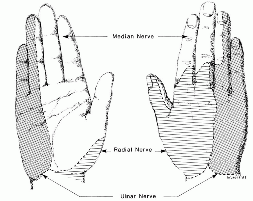
Entrapment neuropathy of the ulnar nerve usually presents with dysesthesias in the sensory distribution of the nerve, often lancinating in quality (Fig. 9.1). Hypesthesia
can follow, again in the ulnar distribution. Finally, weakness of ulnar innervated muscles is a prodrome to atrophy. The latter can lead to the classic claw deformity in the hand. However, the clinical presentation of ulnar nerve entrapment is notoriously variable. Insidiously progressive atrophy can occur with little discomfort and even little sensory loss, the so-called tardy ulnar palsy. More typically there is neuritic discomfort.


FIGURE 9.1. The sensory distribution of the three nerves that innervate the hand.

The ulnar nerve is at risk for entrapment at two sites distal to the shoulder: the elbow and wrist. In the former site, it is superficial at the condylar groove (in the vernacular, the “funny bone”) and further at risk as it passes around the ulna deep to the flexor carpi ulnaris in the “cubital tunnel.” The orientation of the nerve in this tunnel is dynamic; it flattens and stretches with elbow flexion to accommodate the 1-cm increase in the circumference of the posterior elbow capsule. Tinel’s sign can help one gain some confidence that the neuritic symptoms in the distribution of the ulnar nerve actually reflect entrapment about the elbow. Subluxation of the nerve (that it can roll over the medial epicondyle) is nonspecific. Electrodiagnostic studies at this site are highly variable and often not convincing. However, if one can eliminate the two other sites that can mimic this presentation, cervical radiculopathy or entrapment at the wrist, conservative therapy at the elbow is the treatment of choice. Padded splints that buffer the elbow and impede full flexion
are commonly used. Nearly all patients experience complete long-term remission without recourse to surgery.7


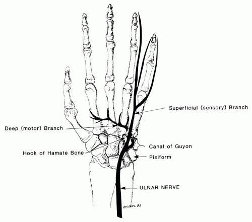
FIGURE 9.2. The anatomy of the ulnar nerve as it courses into the hand.

At the wrist, the ulna nerve is superficial to the flexor retinaculum between the pisiform and the hook of the hamate deep to the superficial volar carpal ligament in a 4-cm conduit called Guyon’s canal (Fig. 9.2). Except for such systemic diseases as rheumatoid arthritis, the most common cause of ulnar entrapment in Guyon’s canal is a ganglion. Lipomas, anomalous muscles, and fractures are other causes. Occupational syndromes involving the ulnar nerve at the wrist are rare. Cyclists often experience ulnar dysesthesias, presumably because of their prolonged posturing; however, they rapidly recover when a normal posture is assumed. The “hypothenar hammer syndrome” is a consequence of repetitive blunt trauma to the hypothenar eminence, usually from using the hand as a hatchet. However, thrombosis and ischemia of the ulnar side of the hand are of greater concern than ulna neuropathy.


Median Nerve Entrapments

The median nerve is susceptible to entrapment as it enters the forearm lying between the two heads of the pronator teres. The diagnosis is suspected when symptoms
indicate CTS, yet Tinel’s sign localizes proximally, and later there is compromise in the power of the long finger flexors. The diagnosis is occasionally confirmed electrodiagnostically but with considerable difficulty. CTS is excluded electrodiagnostically. The diagnosis is aided if pronation against resistance leads to symptoms. This is a rare cause of median neuropathy; the last patient I saw with this disorder was a bass player who experienced the symptoms when he played by picking the strings rather than bowing. Treatment is to restrain forceful pronation so that the pronator muscle involutes.


Carpal Tunnel Syndrome

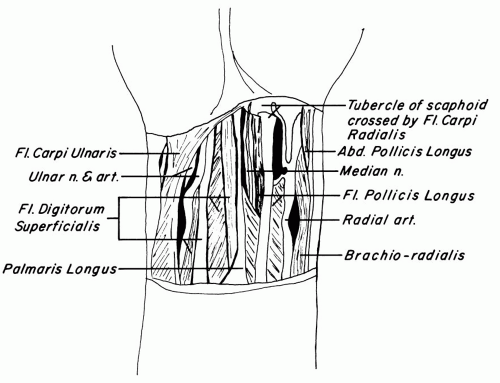
The median nerve is more susceptible to entrapment as it traverses the wrist deep to the flexor retinaculum (Fig. 9.3), which results in CTS. In fact, CTS is the most frequently encountered entrapment neuropathy, but it is still rare. The experience of the Mayo Clinic in caring for its Olmstead County catchment area suggests that the prevalence approximates 99 cases per 100,000 people per year.8 In other words, if you observe 1000 adults for 1 year, you would anticipate one adult to develop CTS. And even that one adult may not come to surgery. In fact, if you restrict the epidemiologic case definition to idiopathic CTS, the incidence is closer to 1 case per 2000 people per year.9 This restricted definition excludes patients with conditions
with known predilection to impair median conductivity at the wrist. CTS is well described as a complication of late pregnancy, hypothyroidism, rheumatoid arthritis, amyloidosis, acromegaly, and diabetes. The nerve can be impinged on with fractures of the wrist or intra-articular inflammatory processes such as tuberculosis. The peripheral neuropathic processes associated with diabetes, amyloidosis, and renal failure have some predilection for the median nerve in the tunnel, rendering these patients at greater risk for CTS but less likely to respond to traditional medical or surgical approaches to this entrapment neuropathy. It is not a manifestation of pyridoxine deficiency; prescribing pyridoxine has an unfavorable risk/benefit ratio.10


FIGURE 9.3. The structures of the volar wrist. Just deep to the palmaris longus tendon is the flexor retinaculum that defines the floor of the carpal tunnel. The median nerve is the only neural structure in the tunnel, accompanied by a number of flexor tendons.

The median nerve at the wrist is peculiarly susceptible to damage in the diabetic patient.11 Impaired conductivity of the median nerve at the wrist is more likely to be bilateral if the patient has diabetes and more likely to afflict the obese, diabetic patient. It is particularly common in diabetic patients who evidence polyneuropathy elsewhere. Impaired median conductivity is present in 14% of patients with diabetes who have no other electrodiagnostic abnormalities and in 30% of patients with polyneuropathy.12 Many diabetic patients with such changes are asymptomatic.13


The Classic Definition of Carpal Tunnel Syndrome

CTS was described in the decade after World War II. The observations from the Mayo Clinic14 were notable in the United States, including those by George Phalen, an alumnus who would pursue his career at the Cleveland Clinic.15 The Mayo Clinic’s case definition is a reflection of the biases that are inherent to observational or ecologic series. For example, Phalen found his sign in approximately 80% of the hands he diagnosed as afflicted with CTS, whereas his colleagues at the Mayo Clinic diagnosed the condition in the absence of Phalen’s sign approximately half the time (Table 9.1). In all likelihood, Phalen’s colleagues were more likely to refer patients with Phalen’s sign to him (referral bias) or he was more adept at eliciting the sign (observer bias). Nonetheless, the Mayo Clinic is offering us a retrospective cohort series; because the Mayo Clinic is the sole provider of medical care in Olmstead County, Minnesota, any person living in the county between 1960 and 1981 who chose to be a patient with hand symptoms that elicited the classic label of CTS are in the Mayo Clinic data set.8 The incidence of approximately one case of idiopathic CTS for every 2000 adults for each year between 1960 and 1981 is not peculiar to Olmstead County. A similar incidence, 0.7 cases per 2000 women per year, was derived from the experience with a cohort of 17,032 English women who were followed from their early 20s for more than 25 years in an effort to discern long-term side effects of oral contraceptive exposure.16 I am belaboring these incidence figures and the classic case definition because they are crucial to understanding the flaws in the contemporary tendency to diagnose CTS in the absence of the referral biases in use for earlier primary care physicians and consultants.

A detailed appreciation of the classic clinical syndrome of CTS is forthcoming from the Mayo Clinic experience. The characteristics of the patients in whom CTS was diagnosed between 1961 and 1980 are presented in Table 9.1. Idiopathic CTS is bilateral in approximately 50% of patients. The clinical presentation is the
prototype for an entrapment neuropathy. The initial presentation is of dysesthesias, even lancinating pain, in the radial side of the hand. If the thumb is involved, the discomfort can extend its length; angina seldom passes the interphalangeal joint. Occasionally the discomfort radiates proximally toward the elbow but seldom further. The dysesthetic sensation often awakens the patient, causing the patient to leave the bed and shake his or her hand. There may be a perception of swelling, even of clumsiness early on without objective correlates. However, with progression, weakness in pinch and even atrophy of the abductor pollicis brevis (Fig. 9.4) are apparent. Further denervation leads to more thenar atrophy and overt thumb weakness. Only approximately half the patients manifest Tinel’s or Phalen’s signs (Fig. 9.5). Furthermore, in this classic series, the Mayo clinicians documented impaired conductivity by electrodiagnostic testing approximately half the time. The clinicians were satisfied with their diagnosis without electrodiagnostic testing the rest of the time and even were satisfied with their diagnosis in the 27% of patients whose electrodiagnostic test results were normal.


TABLE 9.1. CLINICAL FEATURES OF THE PATIENTS WITH DIAGNOSES OF CARPAL TUNNEL SYNDROME AT THE MAYO CLINIC BETWEEN 1961 AND 19808


Total number of patients


1016


Female


798


Male


218


Symmetry


Unilateral


42%


Right


29%


Left


13%


Mean age at diagnosis


Men


50 yr


Women


51 yr


Electrodiagnostic studies


Number studied


505


Percent positive


73


Symptoms


Median paresthesias


100%


Nocturnal paresthesias


71%


Proximal radiation


38%


Percent with signs


Right/left


Positive Tinel’s sign


55/44


Positive Phalen’s sign


55/52


Decreased hand sensation


30/26


Thenar atrophy


20/15


The Contemporary Definition of Carpal Tunnel Syndrome

It is essential to realize that the referral biases that turned Phalen and his contemporaries into legendary observers no longer exist! Today no diagnostician can assume that the signs and symptoms that allowed Mayo clinicians to diagnose CTS in more than 1000 cases (Table 9.1) will pertain to any other experience or to the Mayo Clinic any more. No symptom or sign, short of thenar atrophy, is diagnostic! I can offer this warning with a great deal of confidence in the case of CTS, because there is an objective test that should have abnormal results: the quantification of median conductivity across the wrist. If median conductivity is normal, by definition the patient does not have CTS. I will accept there might be exceptions that relate to the possibility that what is normal for one person may be abnormal for another, but these are truly exceptions. So if one takes electrodiagnostic abnormalities as the gold standard for diagnostic confirmation, as one should, it is possible to test the specificity and sensitivity of the classic signs and symptoms. This was the design of a study performed at the Brigham and Women’s Hospital that was based on patients with various upper extremity symptoms who were referred for electrodiagnostic studies.17 The principal results are presented in Table 9.2. Sensitivity is a property of the test that speaks to the likelihood of false-negative reactions. In other words, if you have 100 people with electrodiagnostically defined CTS, how many will have a particular sign or symptom? Table 9.2 shows that 60% had Tinel’s sign. Specificity is also a property of the test, the likelihood of false-positive reactions. If you have 100 people with upper extremity symptoms and no electrodiagnostic evidence of median neuropathy at the wrist, how many of them will have Tinel’s sign? Table 9.2 shows that 67% had Tinel’s sign. However, seldom do we use a test when we know the subjects do or do not have the disease. We only need tests when we are not sure. Let us say we have 100 people with upper extremity symptoms, only 15 of whom have CTS by electrodiagnostic criteria. Knowing its specificity and sensitivity, how good is Tinel’s sign in finding the 15? Not good. Of any four people with a positive Tinel’s sign, only one is likely to have CTS. That is what is meant by a positive predictive value of 0.25. A negative Tinel’s sign is more useful; only 1 in 10 will have a false-negative reaction (negative predictive value of 0.91). However, for Tinel’s sign to have even these small and inadequate utilities, one needs a referral population in whom some 15% had CTS by electrodiagnostic criteria. If the prior probability of CTS in the referral population increases to approximately 40%, then a positive Tinel’s sign and a positive hand diagram together would have a useful positive predictive value of approximately 70%. If the true prevalence is less, even Tinel’s sign and the diagram would be inadequate for screening.18 It would also be unreliable and invalid for diagnosing CTS in any individual in such a population who is referred to you without some subliminal referral bias, such as that used in Phalen’s practice in the past. Other investigators have quantified the utilities of the classic symptoms and signs of CTS19,20 with results similar to that in Table 9.2. The upshot is that no one should ever make a diagnosis of CTS without electrodiagnostic confirmation any longer!


FIGURE 9.4. The abductor pollicis brevis muscle forms the most lateral aspect of the thenar eminence (arrow). This muscle is innervated only by the median nerve. Therefore, atrophy of the abductor pollicis brevis is diagnostic of median neuropathy and an indication for electrodiagnostic studies to diagnose carpal tunnel syndrome (CTS). If one demonstrates delayed median conduction at the wrist and denervation of the abductor pollicis brevis, surgical division of the flexor retinaculum is advisable.


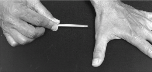
FIGURE 9.5. Tinel’s sign for median entrapment at the wrist is elicited by tapping the palmaris longus tendon over the flexor retinaculum (pointer). If the tap elicits discomfort or dysesthesias that radiate into the thumb, Tinel’s sign is considered positive. Local tenderness at the site of percussion is nonspecific.


TABLE 9.2. BRIGHAM AND WOMEN’S HOSPITAL AND HARVARD MEDICAL SCHOOL ANALYSIS OF THE USE OF CLASSIC SIGNS AND SYMPTOMS OF CARPAL TUNNEL SYNDROME IN 110 PATIENTS WITH ARM PAIN WHO WERE REFERRED FOR ELECTRODIAGNOSTIC TESTING17


Finding (all patients)


Sensitivity


Specificity


Predictive value (assuming 15% prevalence) Positive/negative


Tinel’s sign


0.60


0.67


0.25/0.91


Phalen’s sign


0.75


0.47


0.20/0.91


Hand diagram


0.61


0.71


0.27/0.91


Sensory loss


0.32


0.81


0.23/0.87


Nocturnal symptoms


0.77


0.28


0.16/0.87


Bilateral symptoms


0.61


0.58


0.20/0.89


Similarly, surveillance for arm pain in any population may define the incidence of arm pain with some validity, but not CTS unless there is electrodiagnostic screening.


Fine-Tuning the Electrodiagnosis of Carpal Tunnel Syndrome

With the realization that electrodiagnostic testing is more than a gold standard for the diagnosis of CTS, it is sine qua non, the methodology has been revisited. There has been considerable attention paid to the technique21; trained operators are attentive to the quality of equipment, placement of electrodes, and warmth of the arm. A cool extremity consequent to low ambient temperature or to vasoconstriction from anxiety precipitated by the study will retard conductivity. Electrodiagnostic testing is easy to do poorly!

There has also been a good deal of effort expended in defining the normal range. There is considerable variability in the population. It turns out that much of this can be accounted for by the personal attributes listed in Table 9.3. The implication is that a middle-aged, overweight woman with a square wrist will have slower median conductivity across the wrist than a thin, younger woman whose wrist is more rectangular. Both conductivities can be normal for the women’s habitus and age. Clearly, these factors must be taken into account to interpret electrodiagnostic data on median conductivity either for epidemiologic or diagnostic purposes.22
Even with these advances in the performance and interpretation of electrodiagnostic testing, no one should take away the message that no challenges remain to diagnosing CTS or defining its incidence or prevalence.


TABLE 9.3. PERSONAL ATTRIBUTES THAT MUST BE TAKEN INTO ACCOUNT TO DEFINE NORMAL MEDIAN NERVE CONDUCTIVITY


Epidemiology of median nerve conduction velocities


Variable


Correlation with median conductivity


Age


Negative


Female gender


Negative


Body mass index


Negative


Wrist squareness


Negative


Limitations of Electrodiagnostic Accuracy for Carpal Tunnel Syndrome

The same considerations regarding the utility of symptoms and signs apply to using the gold standard electrodiagnostic test as the diagnostic tool. Its utility will depend on the prior probability of true CTS in the population being studied. Diagnosing CTS is a daunting, if not illusory, challenge consequent to contemporary referral biases (see Chapter 11) that can render the prior probability of the disorder minuscule. Consider the following.

Let us assume that electrodiagnostic testing for CTS is so advanced that the test is 98% specific and 100% sensitive. That means if you have 100 healthy people, the test will fool you twice, and if you have 100 people with CTS, it will not miss any. (Electrodiagnostics will never be such a good test given inherent biologic variability.) Next, let us assume that the incidence rate for CTS derived by analyzing the Mayo Clinic’s retrospective cohort is in the ballpark; if you follow 2000 people for 1 year, one person will develop idiopathic CTS. Finally, let us assume you are, de facto or by chance, involved in surveillance for CTS in a workforce of 3000 for the coming year, perhaps because the company medical department is willing to refer anyone who complains of arm pain to you. Because at least one third of the workforce will be faced with arm pain each year (vide infra), you will see 1000 workers that year. Nerve conductions will be “abnormal” in 20 people with normal nerves. Given what we know of the epidemiology of median nerve conductivity, another one or two people will have abnormal conductivity because of entrapment neuropathy, which might respond to splinting, steroid injection, or surgery. There is no known way to separate the 20 people with normal nerves that happen to be slower from the two people with abnormal nerves. If you try to separate them by virtue of empiric interventions, you will do a disservice to as many as 10 people for every 1 person you may help.

Similar considerations pertain to the population at large. Because of misinformation in the lay press regarding the role of hand usage in its pathogenesis, CTS hangs like an imprecation over the American people. Common sense regarding coping with arm discomfort is dramatically perturbed. The laity can no longer shake off the discomfort of a dysesthetic hand with a facile explanation such as “I must have slept on it funny.” No longer is the rubric, CTS, part of the privileged vocabulary of the subspecialist. Many seek medical attention, often with a chief complaint like “Do I have carpal tunnel syndrome?” American primary care is now primed for CTS so that electrodiagnostic testing and specialist referrals abound. This is a far cry from the biases in patient accrual that nurtured the insights of our clinical forebears, but it raises the same specter of creating prior probabilities that render even electrodiagnostic testing effete.


I suspect that this flood of people with arm pain, few of whom have median neuropathy, into the diagnostic algorithm accounts for the surprising clinical experience of one of the nation’s leading electrodiagnosticians. Radecki was one of the observers who contributed to our understanding of the latency covariates listed in Table 9.3.23 He went so far as to use his sizable referral experience documenting median and ulnar latencies to develop a formula that predicts the median-minus-ulnar palmar latency difference based on the covariates in Table 9.3.24 The formula predicts the result so well that there is no need to perform the tests. What does this mean? It could mean that CTS is a contrivance for reasons we will come to shortly, but I doubt that. More likely, there are very few patients with true CTS in this referral practice so that those few cannot be distinguished from the few whose normal nerves happen to conduct slowly.


Lessons from Community Electrodiagnostic Surveys

From these discussions, one might predict that finding the people in the community who have symptomatic CTS but have not chosen to be patients is an exercise in futility. There are several recent series that concur with me on this point. In a study25 conducted in Maastricht, The Netherlands, 500 people were randomly selected for the query, “Do you wake up at night because of tingling in your fingers?” Fifty people answered in the affirmative, of whom half had “abnormal” electrodiagnostic study results (although not corrected for the covariates). None of the battery of a dozen clinical tests distinguished the half with the “abnormal” nerve conductions from the other half. However, realize that these abnormal tests represent 5% of the total population studied, raising the question of whether they are even disease markers. The reasoning gets quite circular.

A recent British community-based survey of the point prevalence of hand symptoms and their correlation with nerve conduction velocities leads to similar conclusions.26,27 They targeted a random sample of 1000 adults, but only 648 adults responded to their questionnaire. Of these, 212 adults reported hand symptoms, which seemed classic (or nearly so) for CTS in 120. A sample of the 648 respondents submitted to electrodiagnostic testing without correction for the covariates in Table 9.3. The point prevalence of “abnormal” nerve conductions varied between 7% and 16% depending on how “abnormal” was defined. Regardless, the majority of people with “abnormal” test results had no symptoms. And of those with “classic symptoms,” only 18% had abnormal nerve conductions. Yes, the reasoning is circular.

Another similar survey from Lund, Sweden,28 also found that 20% of the population can report symptoms that are classic for CTS. The symptoms are reported as current, occurring within the past month, or lasting 1 month or more. Of this 20% of the population, 20% had “abnormal” median nerve conductivity. That means that 5% of the community would qualify for the CTS label if everyone with symptoms reported to a physician. Do 5% of adults have idiopathic CTS? That is a prevalence of 5%. If the incidence of 1 case per 1000 people per year is relevant, over the course of 50 years for every person with CTS who chose to be
a patient, another one or two people must stay alive at home and suffer in silence. This is not likely.

It is more likely that symptoms, including symptoms that are “classic” for CTS, come and go for most of us regardless of our median conductivity.29 That was the finding of an 11-year community-based longitudinal study.30 This insight even pertains to many patients whose diagnosis of CTS is believed to be certain but who remain “untreated.”31 Most people with “classic” symptoms, signs, and electrodiagnostic findings for CTS are not at risk for persistent symptoms or damage to their hands. They only need some patience to be asymptomatic again.

Only gold members can continue reading. Log In or Register to continue

Stay updated, free articles. Join our Telegram channel

Jul 21, 2016 | Posted by in ORTHOPEDIC | Comments Off on Neurovascular Syndromes of the Extremities

Full access? Get Clinical Tree
Get Clinical Tree app for offline access