Nail Bed and Fingertip Injuries
General Information
Fingertip injuries are the most common trauma in the hand, accounting for up to 50% of emergency room visits for hand care. Traditional treatment was shortening and closure by any possible method. Goals for repair of the injured fingertip include identification and cleaning of injured structures, maintenance of bone length, and meticulous repair of the soft-tissue envelope.
Diagnostic Criteria
Most fingertip injuries occur as low-energy crush injuries such as those produced by closing a door on the digit. Although tissue is disrupted, it is not usually missing (Fig. 1). More powerful crushing injuries from mechanical presses devitalize tissue and may need extensive débridement. Slicing injuries amputate tissue cleanly and may be suitable for replantation. Ripping injuries from power saws combine crushing and slicing and often produce large-tissue defects requiring graft or flap coverage. A brief health assessment should include medical problems, medications, and allergies.
Physical Examination
Identification of the injured structures is critical for repair. Patients present with painful, bleeding digits. After a brief sensory examination, a digital block will relieve pain (anesthesia section). A digital tourniquet controls bleeding and can be fashioned from a 1/2-in. Penrose drain. Physical examination includes an assessment of vascularity in partial amputations. Skin, nail bed, bone, and tendon may be involved, which are often not initially visible on primary inspection. Irrigation with saline and peroxide removes clot and allows visual inspection. X-rays should focus on the injured finger and not the entire hand. Subtle bony injuries are missed when the beam is not centered on the digit. X-rays may also demonstrate soft-tissue defects exposing bone and foreign bodies that require removal.
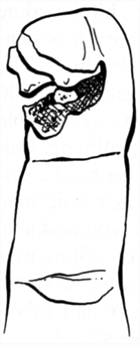 FIG. 1. Incomplete fingertip amputation. |
Treatment
Clear communication with consulting surgeons regarding the nature of the injury, including which tissues are injured, helps guide treatment. Accurate descriptions of bone, nail bed, and pulp injuries facilitate appropriate treatment. Most fingertip injuries can be treated in a well equipped emergency department. Pulp hematomas occur when crushing energy is not strong enough to produce open injuries. Nevertheless, pressure from blood within the pulp produces a painful swollen digit. The cavities between the fibrous septate of the pulp rapidly fill with blood. A nondisplaced tuft fracture is often present. Treatment of these injuries may be purely supportive with analgesics and splints. Alternatively, sterile aspiration of the hematoma and instillation of 0.5 mL bupivacaine relieves pain significantly for 6 to 12 hours. Subungual hematomas of less than 30% of the nail bed also may be treated symptomatically. Drainage of the hematoma using a heated paperclip, 18-gauge needle, or ophthalmic cautery relieves pressure and pain.
Related posts:


Stay updated, free articles. Join our Telegram channel

Full access? Get Clinical Tree


