Abstract
Introduction
Mycetoma is a chronic disease, which is endemic in tropical and subtropical countries. We report a rare case of mycetoma located on the foot of a patient living in a temperate-climate country followed by a short discussion.
Observation
A 50-year-old woman with painless swelling in her left foot. The swelling started after a banal penetrating injury on the sole of her left foot 23 years ago. X-rays images showed multiple osteolytic lesions of the tarsus. The histological analyses identified the fungus madurella mycetomatis. The treatment was radical surgery (amputation). The patient had a tibial prosthesis and recovered an autonomous gait.
Discussion
Mycetoma is a chronic granulomatous inflammatory response often with sinus tract formations due to fungal or bacterial organisms. The infection of the forefoot is quite typical. It is a slowly progressing disease affecting the deep dermis and subcutaneous tissues that can extent to the underlying bones.
Conclusion
If it is not diagnosed early on, mycetoma can cause functional and esthetical impairments.
Résumé
Introduction
Le mycétome est une pathologie infectieuse chronique endémique dans les pays tropicaux, mais rare dans les climats tempérés. Le but de ce travail est de rapporter un cas de localisation au pied, et de rappeler cette affection rarissime, souvent source de difficultés thérapeutiques et de handicap.
Observation
Mme I.D., âgée de 50 ans, consulte pour un pied douloureux, siège de nodules évoluant depuis 23 ans, et survenus dans les suites d’une blessure au niveau de la voute plantaire gauche. La radiographie du pied montre une lyse osseuse du tarse antérieur et du médiotarse. La biopsie osseuse révèle la présence de madurella mycétomatis, relevant d’un traitement chirurgical radical à type d’amputation de la jambe. Une prothèse tibiale a été réalisée avec une récupération de l’autonomie de la marche.
Discussion
Les mycétomes constituent des pseudo-tumeurs inflammatoires, souvent polyfistulisées dues à des champignons ou des bactéries. Ils sont connus par leur localisation podale élective. L’évolution lente et progressive des lésions aux téguments et parties molles finit souvent par une atteinte secondaire du squelette sous-jacent.
Conclusion
Le pied de Madura, non diagnostiqué précocement, peut être à l’origine d’un préjudice fonctionnel et esthétique.
1
English version
1.1
Introduction
Mycetoma is a chronic subcutaneous infection caused by actinomycetes or fungi. This infection results in a granulomatous inflammatory response in the deep dermis and subcutaneous tissue, which can extend to the underlying bone. Mycetoma is characterized by the formation of grains containing aggregates of the causative organisms that may be discharged onto the skin surface through multiple sinuses. It is most commonly seen in tropical and subtropical regions where it can be endemic . The body parts commonly affected by mycetoma are the feet or lower legs with infection of the dorsal aspect of the forefoot being typical. We present here a case of mycetoma infection.
1.2
Case report
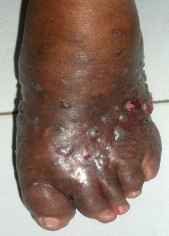
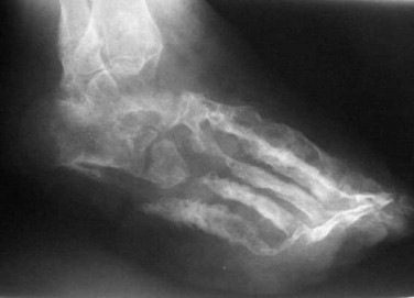
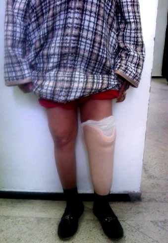
A 50-year-old woman, without any significant medical history and no records of travelling to tropical areas, comes to the consultation with a painless swelling in her left foot. The swelling first occurred, after an injury to the sole of her left foot and has been getting worse every since for the past 23 years. Upon the physical examination we find several sinus tract formations and a large swollen indurate mass palpable on the top of her left foot ( Fig. 1 ). Standard X-rays of her foot showed multiple osteolytic lesions, erosions of the tarsus and soft-tissue swelling ( Fig. 2 ). The histological analysis on the bone biopsy identified a madurella mycetomatis fungal infection ( Fig. 3 ). Due to the extensive bone lesions radical surgery was necessary with amputation below the knee. A prosthesis was designed for this patient and she regained gait autonomy ( Fig. 4 ). A post-operative control conducted six months after surgery did not find any recurrence.


1.3
Discussion
In tropical countries, mycetoma is a real public health issue, but this pathology is quite rare in Morocco . Mycetoma is a chronic granulomatous inflammatory response involving bacteria or fungi that triggers the formation of grains containing aggregates of the causative organisms that may be discharged onto the skin surface through multiple sinuses. It is an infection of the deep subcutaneous soft tissues and bones; this is a severe affection that can develop over several years. It commonly affects young adults aged 20 to 40 years, predominantly males, living in rural areas.
Usually the infection is contracted through a puncture wound in the feet, especially in persons walking barefoot (Madura foot account for 70% of all mycetoma cases). Other localizations include the legs, hands and arms.
This chronic subcutaneous infection can be caused by bacteria or fungi. Mycetoma caused by microaerophilic actinomycetes is termed actinomycetoma, and mycetoma caused by true fungi is called eumycetoma . The next step is to differentiate them through histological skin biopsies to identify the causal agents, if the grains are black or white it is a fungal infection (eumycetoma) if the grains are white, yellow or red it is a bacterial infection (actinomycetoma) . Once the mycetoma is diagnosed, it is essential to look for bone lesions by additional imaging. On X-rays images, bone destruction is visible through multiple and non-specific osteolytic lesions (geodes, cavities, reactive sclerosis). In ultrasound imaging single or multiple thick-walled cavities with hyperreflective echoes and no acoustic enhancement are always observed with mycetoma often aggregated at the lower part of the swollen mass. Nowadays the best definition can be obtained with CT-scan or MRI imaging providing differential diagnoses of the swelling and a better assessment of the degree of bone and soft-tissue involvement . In the treatment of mycetoma medicines and surgery should be combined . The medical treatment consists of antibiotic therapy (cotrimoxazole, amikacin or minocycline) or antifungal therapy (ketoconazole or itraconazole) and should be combined with surgery that should be as thorough as possible. In spite of these treatments, disease recurrence is quite common (50% of the cases). In case of amputation, post-surgical rehabilitation care is essential to provide the patient with proper prosthesis, fittings to the right level of amputation and finally work on balance, posture and gait.
1.4
Conclusion
Mycetoma is a common infection in tropical and subtropical regions (areas with annual rainfall greater than 2500 m). Our case report is a good reminder that this disease can also affect persons living in other areas. Therefore mycetoma should be thought about in differential diagnoses in a chronically swollen and painful foot to avoid, as shown in our case report, a delayed diagnosis leading to functional and esthetical impairments.
Conflicts of interest statement
None.
Stay updated, free articles. Join our Telegram channel

Full access? Get Clinical Tree


