Musculoskeletal/Sports/Orthopedics


Musculoskeletal/Sports/Orthopedics


LIMB JOINT PRIMARY MOVERS


Motion (ROM in degrees)


Muscles


Nerves


Roots


Shoulder flexion (180)


Anterior deltoid


Coracobrachialis


Axillary


Musculocutaneous


C5, C6


C6, C7


Shoulder extension (45)


Latissimus dorsi


Teres major


Posterior deltoid


Thoracodorsal


Inferior subscapular


Axillary


C6, C7, C8


C5, C6, C7


C5, C6


Shoulder abduction (180)


Middle deltoid


Supraspinatus


Axillary


Suprascapular


C5, C6


C5, C6


Shoulder adduction (40)


Pectoralis major


Latissimus dorsi


Med + lat pectoral


Thoracodorsal


C5-T1


C6, C7, C8


Shoulder external rotation (90a)


Infraspinatus


Teres minor


Suprascapular


Axillary


C5, C6


C5, C6


Shoulder internal rotation (80a)


Subscapularis


Pectoralis major


Latissimus dorsi


Teres major


Sup + inf subscapular


Med + lat pectoral


Thoracodorsal


Inferior


subscapular


C5, C6


C5-T1


C6, C7, C8


C5, C6, C7


Shoulder shrug


Trapezius


Levator scapulae


Spinal accessory (CN XI)


C3, C4 ± dorsal scapular (C5)


Elbow flexion (150)


Biceps brachii


Brachialis


Brachioradialis


Musculocutaneous


Musculocutaneous


Radial


C5, C6


C5, C6


C5, C6


Elbow extension


Triceps brachii


Radial


C6, C7, C8


Forearm supination (80)


Supinator


Biceps brachii


Posterior interosseous


Musculocutaneous


C5, C6, C7


C5, C6


Forearm pronation (80)


Pronator teres


Pronator quadratus


Median


Anterior interosseous


C6, C7


C8, T1


Wrist flexion (80)


Flexor carpi radialis


Flexor carpi ulnaris


Median


Ulnar


C6, C7, C8


C7, C8, T1


Wrist extension (70)


Ext carpi rad longus


Ext carpi rad brevis


Ext carpi ulnaris


Radial


Radial


Posterior interosseous


C6, C7


C6, C7


C7, C8


MCP flexion (90)


Lumbricals


Dors + palm interossei


Median, ulnar


Ulnar


C8, T1


C8, T1


PIP flexion (100)


Flexor digitorum sup


Flexor digitorum prof


Median


Median, ulnar


C7-T1


C7, C8, T1


DIP flexion (90)


Flexor digitorum prof


Median, ulnar


C7, C8, T1


MCP, finger extension


Extensor digitorum


Extensor indicis


Extensor digiti min


Posterior interosseous


Posterior interosseous


Posterior interosseous


C7, C8


C7, C8


C7, C8


Finger abduction (20)


Dorsal interossei


Abductor digiti min


Ulnar


Ulnar


C8, T1


C8, T1


Finger adduction


Palmar interossei


Ulnar


C8, T1


Thumb flexion


Flexor pollicis brevis


Flexor pollicis longus


Median, ulnar


Anterior interosseus


C8, T1


C7, C8, T1


Thumb extension


EPB


Extensor pollicis


longus


Posterior interosseous


Posterior interosseous


C7, C8


C7, C8


Thumb abduction


Abd pollicis longus


Abd pollicis brevis


Posterior interosseous


Median


C7, C8


C8, T1


Thumb adduction


Adductor pollicis


Ulnar


C8, T1


Hip flexion (120)


Iliopsoas


Femoral


L2, L3, L4


Hip extension (30)


Gluteus maximus


Inferior gluteal


L5, S1, S2


Hip abduction (40)


Gluteus medius


Gluteus minimus


Superior gluteal


Superior gluteal


L4, L5, S1


L4, L5, S1


Hip adduction (20)


Adductor longus


Adductor magnus


Obturator


Obturator, sciatic


L2, L3, L4


L2, L3, L4,


L5, S1


Hip external rotation (45)


Obturator int + ext


Quadratus femoris


Piriformis


Sup + inf gemelli


Glut max (postfibers)


n. obt int, obturator


n. quadratus femoris


n. piriformis


n. obt int, n. quad fem


Inferior gluteal


L3-S2


L2, L3, L4


S1, S2


L4-S2


L5, S1, S2


Hip internal rotation (45)


Gluteus minimus


Gluteus medius


Tensor fasciae latae


Superior gluteal


Superior gluteal


Superior gluteal


L4, L5, S1


L4, L5, S1


L4, L5, S1


Knee flexion (135)


Semitendinosus


Semimembranosus


Biceps femoris


Tibial div. of sciatic


Tibial div. of sciatic


Tib + per div. sciatic


L5, S1, S2


L4, L5-S2


L5, S1, S2


Knee extension


Quadriceps femoris


Femoral


L2, L3, L4


Ankle dorsiflexion (20)


Tibialis anterior


Deep peroneal


L4, L5, S1


Ankle plantarflexion (45)


Gastrocnemius


Soleus


Tibial


Tibial


L5, S1, S2


L5, S1, S2


Ankle inversion (35)


Tibialis posterior


Tibial


L4, L5, S1


Ankle eversion (25)


Peroneus longus


Peroneus brevis


Superficial peroneal


Superficial peroneal


L4, L5, S1


L4, L5, S1


Toe extension


Extensor hallucis longus


Extensor digitorum brevis


Deep peroneal


Deep peroneal


L4, L5, S1


L5, S1


a Shoulder IR/ER varies with elevation of the arm.


CN, cranial nerve; IR, internal rotation; PIP, proximal interphalangeal.


For ROM , 0° is anatomic position. Please note that there is no absolute consensus regarding which muscles are the primary movers of joints or for the root innervations of muscles.


TREATMENT OF SELECTED MSK CONDITIONS


Upper Limb

AC Sprains/TearsAC injuries may be seen with falls on the adducted shoulder. A type I (Rockwood classification) injury is a nondisplaced sprain of the AC ligament, manifested by local tenderness w/o
anatomic deformity. A type II injury (see Fig. 7-1) involves an AC tear and CC ligament sprain, but the CC interspace is intact. Treatment for type I or II injuries includes an arm sling, ice, analgesics, and progressive ROM exercises. An unstable type II injury may require arm sling use for 2 to 4 weeks. Sports activities can be resumed when full painless ROM is achieved and deltoid strength is near-baseline. Type III to VI lesions involve rupture of the AC and CC ligaments with varying displacements of the clavicle. These require orthopedic consultation for potential ORIF, although many separations may be followed conservatively with several weeks of sling-and-swathe immobilization, followed by long-term therapy.


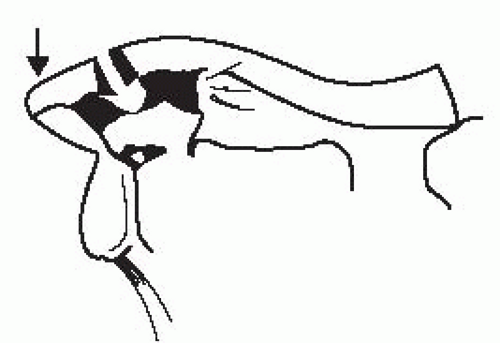
Figure 7-1 Type II injury.

Adapted from Rockwood CA, ed. Rockwood & Green’s Fractures in Adults. 3rd ed. Philadelphia, PA: JB Lippincott; 1988.

ACJ OAOA is a very common cause of ACJ pain, especially in the elderly. The presence of ACJ tenderness and pain with cross body abduction suggests ACJ OA. Radiologic studies such as x-rays and US evaluation can help confirm the diagnosis. Treatment includes topical or oral analgesics, PT, injections, and surgery if refractory to conservative care. Traditional injection techniques have proven to be inaccurate; therefore, fluoroscopic or US-guided injections are preferred.1

ACJ OA (Rotator Cuff Tendinitis/Shoulder Impingement Syndrome) – Predisposing and causative factors include acromion shape and repetitive overhead activities (i.e., throwing, racquet sports, and swimming). Pain and aches are often worse at night and can be aggravated by overhead activities. Shoulder flexion and abduction may be limited.

A painful arc (Fig. 7-2) may be present at about 70° to 110° on passive arm abduction. Neer’s test (Fig. 7-3) and Hawkins test evaluate for shoulder impingement. In Neer’s test, the examiner fixes the scapula with one hand and elevates the subject’s arm with the other hand. Pain indicates a positive test. Hawkins test is performed by abducting the subject’s arm to 90° with the elbow flexed, then internally rotating the shoulder. Hawkins test can also be performed in the
scapular plane. In the drop arm test, the arm is passively elevated to 90° in abduction and the patient is asked to hold the arm in position and then slowly lower the arm to the side. The inability to slowly lower the arm or having severe pain when attempting to do so may be indicative of a severe or complete tear of the rotator cuff pathology.


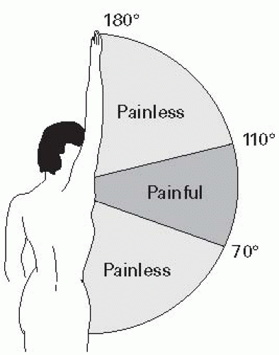
Figure 7-2 Painful arc.


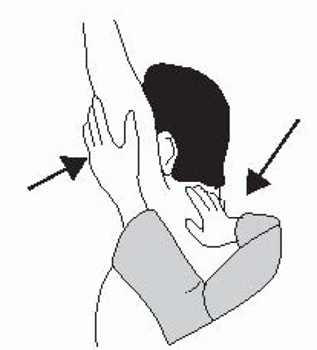
Figure 7-3 Neer’s test.

The painful shoulder should initially be rested until pain and swelling subside. Ice and NSAIDs may be helpful. Overhead activities should be avoided. PT can institute gentle stretching to preserve ROM and isometric strengthening. A steroid injection into the subacromial space may relieve pain and improve motion if the above measures fail. A repeat injection should be avoided in patients with <2 months of pain relief following the first injection. Unless your clinical diagnosis is unchanged, repeat subacromial injection with US guidance to ensure accurate medication placement may be considered for additional diagnostic and therapeutic purposes.2,3 Exercises should progress until strength and ROM are restored. Surgery is an option if several months of conservative treatment/steroid injections fail to resolve the symptoms (or for complete tears). An acromioplasty, the most common procedure, involves acromial shaving to increase the space around the inflamed tendon. The tendon may also be debrided. Several months may be required to regain full strength after surgery.

Anterior Shoulder Dislocation – Anterior dislocations are more common than posterior dislocations. Complications include axillary nerve injury, recurrent dislocations, and rotator cuff tears (especially in older patients). A Bankart lesion (Fig. 7-4) is an avulsion of the anteroinferior glenoid labrum and capsule from the glenoid rim and is felt to be a primary etiologic factor in recurrent dislocations. A Hill-Sachs lesion is a compression fracture of the humeral head when the posterolateral aspect of the humeral head compresses against the anterior glenoid rim. Age at initial dislocation is prognostic for recurrence: teens/young adults have significantly higher redislocation rates (said to approach 90%) than older patients (said to be ≈10% to 15% for patients >40 years of age).

Various techniques exist for acute reduction, including the modified Stimson technique, where the patient lies prone with a wrist weight (i.e., 5 to 10 lbs) on the affected arm as it hangs over the side of the table. Reduction is achieved over 15 to 20 minutes as the shoulder muscles relax.


Figure 7-4 Bankart lesion.

A newer technique termed FARES has been published and appears to be superior to the Hippocratic and Kocher methods, but was not compared with the
Stimson technique. This method is performed with the patient supine and longitudinal traction is applied as the shoulder is slowly abducted.4 Neurovascular status should be checked before and after attempted relocation.

There is no strong evidence to show that immobilization or the duration of immobilization has an effect on the outcome. One option includes bracing in ER, which may reduce the rate of recurrence, though this should be initiated 24 to 48 hours following injury. Early rehabilitation may include icing and sling immobilization for 1 to 3 weeks to allow healing of the capsule. Maintenance of elbow, wrist, and hand ROM is important. Isometric exercises and gentle pendular exercises with the arm in the sling are encouraged, but passive abduction for hygiene is limited to 45° and ER is avoided. The duration of sling use may be shortened in older patients due to the higher risk of frozen shoulder. Once the capsule has healed, shoulder ROM and strengthening are progressed. There is some debate regarding the optimal type and timing of surgery after shoulder dislocation and in shoulder instability.

Adhesive Capsulitis – A syndrome characterized by a progressive painful loss of passive and active glenohumeral ROM that occurs more commonly in females between the ages of 40 and 60 years. Abduction and ER are most affected; internal rotation (IR) is least affected. This condition may be the end result of other conditions that result in prolonged immobility (i.e., bursitis and rotator cuff tendinitis) and has also been associated with other medical conditions (i.e., DM, thyroid dysfunction, and autoimmune diseases). Treatment can consist of an aggressive ROM program, with NSAIDs and heat modalities to improve tolerance. Other techniques include intra-articular steroid injections, brisement (hydrodilation of the capsule), manipulation under anesthesia, and suprascapular nerve blocks. Of these additional options, intra-articular steroid injection has been well studied and appears to improve short-term outcomes. Recovery may take several months to beyond a year.5

Bicipital Tendinitis – This overuse injury can be associated with overhead activities or sports and often coexists with the shoulder impingement syndrome, rotator cuff tears, or labral pathology (i.e., SLAP lesions). Examination often reveals a tender bicipital groove. While palpating this structure, assess for instability/subluxation of the bicipital tendon by internally and externally rotating the shoulder. If unstable, the tendon may sublux medially over the lesser tuberosity and a clunk or snap may be appreciated. Speed’s test (Fig. 7-5) is performed by elevating the subject’s arm to 90° with the elbow extended and palm upward, then having the patient attempt forward flexion of the arm against resistance. Pain in the bicipital groove is indicative of a positive test. Treatment includes NSAIDs, activity modification, and progressive exercise
program, which may include the use of modalities such as heat and postactivity icing. Local corticosteroid injection may be used in refractory cases and US guidance may help increase your accuracy of performing injections into the tendon sheath.6 Scapular Winging – Medial scapular winging (Fig. 7-6) is caused by weakness of the serratus anterior (long thoracic nerve). It is elicited by having the patient push against a wall and using resisted forward flexion or resisted scapular protraction.


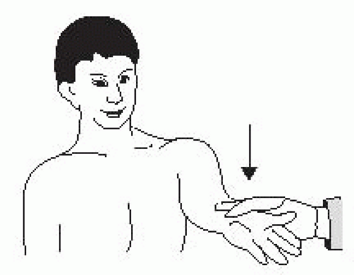
Figure 7-5 Speed’s test.


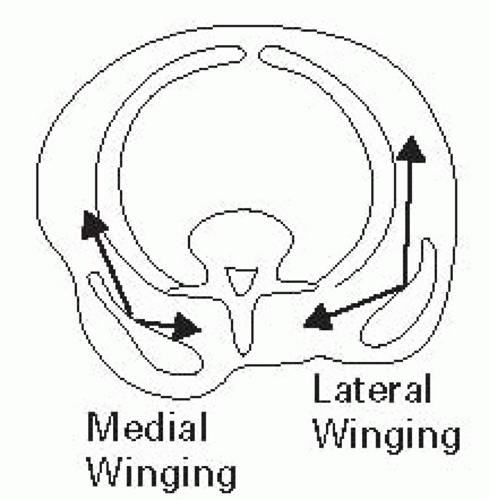
Figure 7-6 Scapular winging.

Lateral winging is caused by weakness of the trapezius muscle (CN XI) and is elicited by shoulder abduction.

Golfer’s Elbow (medial epicondylitis) – An overuse syndrome of the tendinous origin of the flexor-pronator mass and medial collateral ligament of the elbow. The initial treatment is RICE and NSAIDs. Stretching the elbow during the painful period is important. Once pain and inflammation subside, strengthening exercises are started (important groups include the wrist flexors/extensors, wrist radial deviators, forearm pronator/supinators, and elbow flexor/extensors). Injection of local steroids into the area of max tenderness can also be considered, with care taken not to injure the ulnar nerve. A tennis elbow counterforce strap may be helpful.

Tennis Elbow (lateral epicondylitis) – An extensor tendinopathy, especially of the ECRB. The initial treatment is relative rest, NSAIDs, and heat or cold modalities. Wrist extensor stretching and strengthening should be initiated when tolerated. Conservative measures are usually effective, but recurrences are common. A tennis elbow strap worn circumferentially around the forearm just distal to the elbow may be helpful and a wrist splint may be considered to rest the common wrist extensor tendons. Modifications to the racquet include a larger racquet grip and head and lesser string tension. A corticosteroid injection into the area of max tenderness may be indicated if conservative treatment fails. Treatment with PRP or autologous whole blood has been shown to be more effective than corticosteroids in those patients who have failed conservative treatment.7,8 No more than three injections should be given at intervals of 5 days to 1 week. Surgical fasciotomy or fixation of the conjoined tendon may be considered if the above measures fail.

Only gold members can continue reading. Log In or Register to continue

Stay updated, free articles. Join our Telegram channel

Jun 19, 2016 | Posted by in PHYSICAL MEDICINE & REHABILITATION | Comments Off on Musculoskeletal/Sports/Orthopedics

Full access? Get Clinical Tree

Get Clinical Tree app for offline access