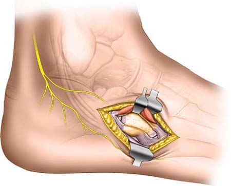
 Midfoot: Approach to the Cuboid
The midfoot consists of the navicular, cuboid and cuneiform bones, their joints, and the four powerful muscles that insert into the midfoot that are responsible for controlling inversion and eversion of the foot. The muscles are the tibialis anterior, which inserts into the medial surface and undersurface of the medial cuneiform bone and into the adjoining part of the base of the first metatarsal bone; the peroneus longus, which inserts into the lateral side of the medial cuneiform bone; the peroneus brevis, which inserts into the base of the lateral side of the fifth metatarsal bone; and the tibialis posterior, which inserts into the tuberosity of the navicular bone, the inferior surface of the medial cuneiform, the intermediate cuneiform, and bases of the second, third, and fourth metatarsal bones.
Proximally, the midfoot begins at the calcaneal cuboid joint laterally and the talonavicular joint medially. Distally, it ends at the joint between the cuboid and the lateral metatarsals laterally and the joint between the cuneiforms and the medial metatarsals medially.
All bones of the midfoot are superficial and can be approached directly by dorsal, medial, and lateral approaches. The middle part of the foot is the target of various specialized procedures for the treatment of muscle imbalance, mobile flat foot, accessory navicular bone, as well as fracture care.
This approach is used mainly for the treatment of cuboid fractures. These injuries are frequently associated with other midfoot and hindfoot fractures; therefore this approach is often combined with other surgical approaches. Careful assessment of the skin and associated soft-tissue injuries is essential before considering surgery. Procedures may have to be delayed to allow swelling to subside and soft-tissue injuries to heal.
Position of the Patient
Place the patient on the operating table in the lateral position (see Fig. 11-1). Ensure that all bony prominences are well padded and that the patient is stabilized, using a bean bag or kidney rests. It is best to have the under leg flexed at the knee with the top leg more extended if fluoroscopy is needed. After exsanguination, apply a tourniquet to the mid-thigh.
Stay updated, free articles. Join our Telegram channel

Full access? Get Clinical Tree


