 Medial Approach to the Sustentaculum Tali
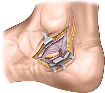
This approach to the hindfoot provides exposure to the sustentaculum tali as well as the flexor tendons on the medial side of the foot. The indications for its use include sustentacular fractures, the release of tendons, and treatment of inflammatory conditions.
Position of the Patient
Place the patient supine on the operating table with a bump underneath the opposite side hip. This will roll the foot into external rotation (see Fig. 1-1). After exsanguination, apply a tourniquet to the mid-thigh.
Landmarks and Incision
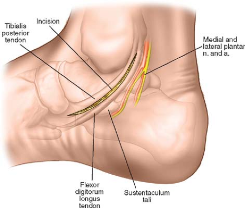
The medial malleolus is the bulbous, subcutaneous distal end of the medial surface of the tibia. It is the best landmark for this incision. Palpate the pulse of the posterior tibial artery immediately posterior and distal to the medial malleolus before inflating the tourniquet. Mark its position on the skin with an indelible marker. Next, palpate the bony tip of the navicular on the medial side of the foot, just distal and plantarward from the tip of the medial malleolus. Finally, palpate the sustentaculum tali. It is felt as a bony resistance deep to the tibialis posterior and flexor digitorum longus immediately distal to the tip of the medial malleolus.
Make an 8-cm curved incision on the medial aspect of the hindfoot. Begin the incision starting at the bony tip of the navicular (Fig. 24-1). The incision should be distal to the medial malleolus and overlie the bony prominence of the sustentaculum tali. Take great care to cut only the skin, as the neurovascular bundle is very superficial at this point.
Internervous Plane
This approach does not use an internervous plane. All of the muscles seen receive their nerve supply well proximal to the approach and therefore are not denervated by it.
Stay updated, free articles. Join our Telegram channel

Full access? Get Clinical Tree


