 Medial Approach to the First Metatarsal Bone for Excision of the Medial Sesamoid Bone
This surgical approach is used almost exclusively for excision of the medial (tibial) sesamoid bone.
Two structures are at risk during this surgical procedure. The medial plantar sensory nerve lies just dorsal to the medial sesamoid, and must be identified and preserved to avoid postoperative anesthesia in weight-bearing areas. The flexor hallucis longus tendon is also at risk during excision of the medial sesamoid bone.
Position of Patient
Place the patient supine on the operating table. After exsanguination, place a tourniquet on the middle of the thigh. Alternatively, use a soft rubber bandage to exsanguinate the foot, then wrap the leg tightly just above the ankle (see Fig. 1-1).
Landmarks and Incision
Palpate the head of the first metatarsal bone and the metatarsophalangeal joint on the ball of the foot and along its medial border.
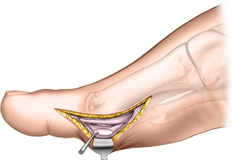
Make a 3- to 4-cm longitudinal incision on the medial aspect of the foot. Begin just distal to the metatarsophalangeal joint of the hallux overlying the plantar border of the joint. Extend the incision proximally to follow the plantar border of the first metatarsal bone (Fig. 39-1).
Internervous Plane
There is no true internervous plane. The two muscles encountered during the approach—the abductor hallucis and the flexor hallucis longus—receive their nerve supply well proximal to the field of dissection, therefore neither muscle can be denervated by this procedure.
Stay updated, free articles. Join our Telegram channel

Full access? Get Clinical Tree


