Lower Extremity Considerations: Hip
Michael N. Kang
Daniel J. Berry
William J. Maloney III
Introduction
There has been much debate whether OA of the hip is a primary or secondary process. A pre-existing condition such as hip dysplasia, slipped capital femoral epiphysis, Perthes disease, and previous trauma are well-recognized processes that lead to osteoarthritis (OA).1 Ganz and colleagues have proposed a different primary process referred to as femoroacetabular impingement. Regardless of the underlying etiology, the end result of OA is loss of articular cartilage and subsequent joint deformity. Clinically, the patient experiences progressively increasing frequency and severity of pain with loss of range of motion. First-line therapy includes medications such as acetaminophen and non-steroidal anti-inflammatory medications, nutritional supplements such as glucosamine and chondroitin sulfate, activity modification, weight loss, physical therapy, and a walking aid such as a cane.
This chapter focuses on the surgical considerations for treatment of OA of the hip—primary and secondary. Surgical options depend on the diagnosis, severity of arthritis, patient age and activity level, patient occupation, patient medical health, and patient expectations. Surgical options are grouped into five main categories. Hip arthroscopy is utilized for the pre-arthritic hip with labral lesions. Surgical débridement and reshaping of the femoral head and neck through open and arthroscopic approaches is used to treat femoroacetabular impingement in the early stages of arthritis. Hip arthrodesis or fusion is indicated in the very young patient with end stage arthritis. Osteotomy is an option mainly in patients who have hip dysplasia. An osteotomy realigns the acetabulum, normalizing the forces that are transmitted through the hip joint and subsequently relieve pain. Finally, hip joint arthroplasty involves replacing all or parts of the diseased joint with artificial components. Each category will be discussed individually.
Arthroscopy
The development of arthroscopic surgery of the hip has been slower to evolve in comparison to other joints such as the knee or shoulder due to more complex anatomic constraints as well as the fact that conditions of the hip like labral pathology can go unrecognized and untreated. As a less invasive tool to diagnose and treat hip pathology, the indications for hip arthroscopy most commonly include labral tears, capsular laxity, chondral injury, ligamentum teres avulsions, and removal of loose bodies. Less commonly, they can include management of osteonecrosis, inflammatory synovial processes, infection, and possibly early to mild OA.2,3
Advanced imaging studies such as computed tomography (CT), magnetic resonance imaging (MRI), and magnetic resonance arthrography (MRA) have improved the ability to diagnose bony and soft tissue pathology about the hip. However, Edwards and colleagues4 reported that MRI was relatively poor in the diagnosis of chondral fibrillation or defects under 1 cm. Furthermore, MRI did not reliably diagnose loose bodies and labral tears. Gadolinium-enhanced MRA has improved diagnostic sensitivity and accuracy.5 The gold standard of diagnosis remains visualization via arthroscopy.
Keeney and colleagues6 evaluated the effectiveness of MRA as a diagnostic tool. In 102 hips, MRA was obtained in order to confirm the diagnosis of labral pathology as well as exclude other conditions that could contribute to hip pain. MRA was able to diagnose 71 out of 102 hips with labral pathology. The sensitivity is 71%, while the positive predictive value is 93%. Articular pathology was also assessed, and MRA demonstrated a sensitivity of 47%, specificity of 89%, and a positive predictive value of 84%. The authors conclude that MRA is an effective tool to diagnose labral pathology; however, a negative study does not exclude intra-articular pathology that can be treated arthroscopically.
For the patient 1) who complains of mechanical symptoms in the hip, 2) has physical exam consistent with an intra-articular process, and 3) has supporting imaging studies, arthroscopic débridement of labral tears has shown to be effective in nearly 90% by Philippon et al.7 It is important to differentiate an intra-articular process versus external pathology, such as psoas tendon irritation over the iliopectineal eminence or femoral head.8 McCarthy and colleagues9 performed 436 hip arthroscopies and determined that 55% had labral tears. In the labral tear group, 73% were found to have associated chondral injury, and chondral injury was more prevalent in older patients. The authors hypothesize that the altered biomechanics of the hip joint lead to labral tears and subsequent degenerative changes in the cartilage.
Femoro-Acetabular Impingement
Femoro-acetabular impingement is a concept that has been championed by Ganz as a cause of secondary OA.10 Abnormal or excessive contact between the proximal femur and acetabular rim leads to lesions in the labrum and chondral surfaces that can result in degenerative changes of the entire hip joint. Initial radiographic work-up may appear relatively normal. Physical examination involves provocative tests to cause the bony impingement. Anterior impingement can be assessed with hip flexion and internal rotation, while posterior impingement can be re-created by extension and external rotation.
Radiographic examination often demonstrates a bony prominence on the anterolateral aspect of the femoral neck as well as possible herniation pits in this region. Other bony abnormalities, such as acetabular retroversion, acetabular protrusion, hip dysplasia, and coxa vara or valga, can also be present. MRA should be routinely obtained in this patient population as there is a high incidence of labral and chondral pathology.11
Two types of impingement have been described: cam impingement and pincer impingement. Cam impingement is caused by an abnormal femoral head with a larger radius being forced into a smaller acetabulum, especially in flexion.12 This produces shear forces that produce abrasion of the cartilage or avulsion of the labrum at the anterosuperior rim. Pincer impingement involves over-coverage anteriorly by the acetabulum leading to impingement and subsequent labral degeneration. Chronic abutment can lead to further ossification of the anterior rim which deepens the acetabulum, worsening this condition. Chronic impingement leads to a “lever” effect and chondral injury in the posteroinferior aspect of the acetabulum.
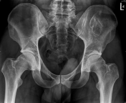 Figure 21A-1A Preoperative radiograph of femoro-acetabular impingement. |
When conservative therapy is inadequate, surgical dislocation of the femoral head via a trochanteric flip osteotomy, as described by the Swiss group, is suggested.13 Surgical treatment includes débridement of the femoral neck to improve offset in order to alleviate the cam impingement. Care must be taken to avoid the retinacular vessels as they enter the superior portion of the femoral neck region. Anterior acetabular bony impingement can also be reduced (pincer impingement). Intra-articular pathology can be treated as well (Fig. 21A-1A,B).
Ganz et al.14 reviewed the surgical treatment in 19 hips with an average follow-up of a minimum of 4 years. The average age of the patients was 36 years. At the latest follow-up, there was significant improvement in the Merle d’Aubigne hip score and pain score. There was an increase in range of motion, though not significant. Thirteen patients had considerable improvement in pain, while two patients had no change. Four patients developed increasing pain in the hip. These four patients and one other went on to total hip arthroplasty. The grade of OA was important in the long-term prognosis of these patients. Of the five hips that underwent conversion to total hip arthroplasty, two hips had grade 2 arthritis, and the other three hips had extensive acetabular cartilage degeneration as well as focal defects in the femoral head.
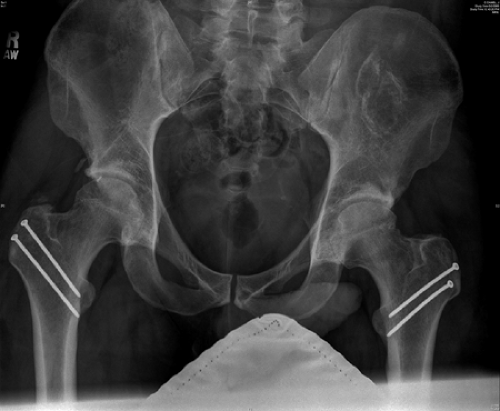 Figure 21A-1B Postoperative radiograph after surgical dislocation and recontouring of femoral neck. |
Other groups have demonstrated similar results. Murphy and colleagues15 describe their experience with débridement of the neck with surgical dislocation in 23 patients. Fifteen patients were satisfied with the procedure.
Seven patients went on to total hip replacement between 6.4 and 9.5 years, while one patient had an arthroscopic procedure for recurrent labral pathology. The Merle d’Aubigne hip score was significantly improved postoperatively in the 15 patients with well-functioning hips. Clohisy and McClure16 describe a different surgical technique to treat anterior impingement. The patients undergo arthroscopic evaluation of the hip joint with debridement of any chondral and labral pathology, followed by a decompression of any bony impingement via an anterior approach. The authors report good results with this technique (Clohisy, unpublished data).
Seven patients went on to total hip replacement between 6.4 and 9.5 years, while one patient had an arthroscopic procedure for recurrent labral pathology. The Merle d’Aubigne hip score was significantly improved postoperatively in the 15 patients with well-functioning hips. Clohisy and McClure16 describe a different surgical technique to treat anterior impingement. The patients undergo arthroscopic evaluation of the hip joint with debridement of any chondral and labral pathology, followed by a decompression of any bony impingement via an anterior approach. The authors report good results with this technique (Clohisy, unpublished data).
Acetabular retroversion has become more commonly diagnosed as a cause of femoroacetabular impingement. Radiographic diagnosis can be performed by the “cross-over and posterior wall” signs.17 Siebenrock et al.18 reported on 29 patients with acetabular retroversion who underwent periacetabular osteotomy to relieve the anterior femoroacetabular impingement. Any intra-articular pathology was also addressed. Twenty-six patients had good to excellent results with significant increase of range of motion. One patient had recurrent anterior impingement, while one had posterior impingement due to overcorrection. In summary, it is important to diagnose the underlying etiology of femoro-acetabular impingement as well as any intra-articular pathology in order to determine the appropriate surgical procedure.
Arthrodesis
Arthrodesis of the hip is a reliable procedure to eliminate pain in young patients (ages 16 to 30) who have prematurely developed end stage arthritis. Due to the successful functional outcomes of total hip arthroplasty (THA) in the older patient, the indications for THA have been stretched to this younger population with the advent of newer, more durable materials. Furthermore, THA is better accepted as it preserves joint mobility and improved gait mechanics, sitting comfort, and sexual function. However, numerous authors have reported on THA failure rates of 33% to 45% in this young patient population.19,20,21,22 The higher failure rate has been attributed to increased activity, leading to increased wear.
A successful hip arthrodesis can provide an active lifestyle that can be later converted to THA.23,24 The indications for hip fusion are young age (< 30 years) with nonin-flammatory, monoarticular, end stage arthritis, especially if any neurologic or muscle (abductor) imbalances exist. Absolute contraindications include active infection, inflammatory arthritis, and radiographic evidence of arthrosis with symptomatic stiffness of the ipsilateral knee, spine, or contralateral hip.25
The position of hip fusion affects outcome. The optimal position for hip fusion is 20° to 30° of hip flexion, 5° to 7° of adduction, and 5° to 10° of external rotation.26 As noted, hip position is important, as too much extension can prevent a comfortable sitting position, while too much flexion can cause increased lumbar lordosis with standing. Discrepancies in abduction or adduction can lead to pelvic obliquity as well as apparent limb length discrepancies. It is recommended that an intraoperative radiograph be performed prior to finalizing position.
The surgical techniques have evolved, however, the principles remain: maximize bone contact, rigid internal fixation, and slightly medialize the hip center. Intra-articular contact with rigid fixation of the ilium to the proximal femur allows for initial stability of the fusion, while permitting mobilization of adjacent joints.27,28,29,30 Early accepted techniques included stripping the abductors and placing a lateral cobra plate contoured to the anatomy of the pelvic brim. Matta et al.28 describe a different technique of anterior plating which spares scarring of the abductors during subsequent conversion to THA. External fixation has been described in the pediatric literature.31,32 In the modern literature, nonunion rates have ranged from 10% to 20%. Nonunions typically require reoperation.
Several groups have reported good results for hip fusion in terms of pain relief and functional ability; however, nearly 70% felt that their activity level was below their respective age group.23,33 Functionally speaking, patients have an asymmetric, arrhythmic gait that is pain free.34,35 The majority of patients lead productive lives without limitations except for activities requiring the extremes of hip flexion. The major long-term complication of hip arthrodesis is ipsilateral knee and lower back arthrosis. Hauge36 reported that 65% of 200 patients developed radiographic evidence of OA of the ipsilateral knee. Callaghan et al.33 had similar results with 60% of the patients experiencing either lower back or ipsilateral knee pain. Furthermore, they noted that there was a trend toward increased arthrosis with malposition of the hip fusion.
A hip fusion, whether it be spontaneous or iatrogenic, can be converted to a THA.24 The primary indications are to relieve symptoms of increasing pain in the lumbar spine, ipsilateral knee, or contralateral hip. It is important to maintain hip abductor function in the index hip fusion and during the subsequent takedown of the hip fusion to permit normal gait. Patients with damage to the abductor muscles will experience a chronic Trendelenburg gait following THA. In addition, adequate tension of the abductors is important during the conversion to THA to diminish the risk of postoperative dislocation. Abductor function can be assessed preoperatively by palpating the contraction of the abductors. Finally, choice of implants (constrained implant) may be affected by a poorly functioning abductor mechanism that may compromise the long-term efficacy and survival of the implants.
Hardinge et al.37 reported on 112 patients converted to THA from either a spontaneous or surgical hip fusion, excluding ankylosing spondylitis. Limb length discrepancies remained in 11.5% of patients, however, only 5% of all patients were dissatisfied with the results. The authors noticed that patients who had fusions prior to skeletal maturity had underdeveloped greater trochanters, and subsequently, abductor function was poor after conversion to THA. Kilgus et al.38 reported that relief of back pain was higher than the relief in ipsilateral knee pain, or contralateral hip or knee pain. The UCLA hip function scores did not improve after THA, reflecting the high level of activity of the patients with a hip fusion. Only 33% of the patients were able to use a less restrictive ambulatory aid.
Caution should be undertaken when performing an arthroplasty of the ipsilateral knee of a fused hip. Garvin et al.39 reported on nine patients who received total knee replacements under a fused hip. Seven patients were available for follow-up; all required at least one postoperative manipulation, and two were unable to flex to at least 90 degrees. The overall complication rate was 65%. Consideration for the takedown of the hip fusion and conversion to a THA prior to knee replacement must be undertaken.
In summary, hip fusion is a reliable operation to eliminate hip pain in the very young patient. Patients can be converted to THA to obtain pain relief in adjacent joints, correct leg length discrepancies, and improve hip mobility. It is important to maintain abductor function during conversion to THA. Although patients are satisfied after conversion to THA, hip function scores do not improve significantly.
Osteotomy
Osteotomies around the hip joint are joint-preserving procedures that are an acceptable alternative to joint replacement surgery in younger patients for the appropriate diagnoses.40 The underlying diagnoses in whom one would consider an osteotomy include young patients with secondary OA from hip dysplasia and residual deformities from childhood conditions such as Perthes disease and Slipped Capital Femoral Epiphysis (SCFE). Other indications for osteotomies include partial osteonecrosis of the femoral head and femoral neck fracture nonunions; however, these fall outside of the scope of this chapter.
In the 1960s, the introduction of THA markedly diminished the utilization of osteotomy for OA. However, as long-term studies demonstrated that THA lacked durability past 15 to 20 years, especially in younger patients, hip osteotomies have become more attractive as they have the potential to provide excellent results with good long-term durability in the appropriately selected patient. The goal of an osteotomy is to relieve pain by redirecting the distribution of load and changing the stress gradients, but there is little change in the actual joint loads.41 Hip osteotomies can provide durable pain relief by one or a combination of the following mechanisms: 1) improvement in joint congruity leading to increased joint contact area and decreased joint contact stress; 2) improvement in hip biomechanics decreasing joint contact forces; 3) rotation of intact articular cartilage into the weight-bearing dome, thus loading more normal cartilage; and 4) reduction in joint subluxation decreasing shear stresses on the articular cartilage.
Osteotomies around the hip joint can be performed on the pelvic or the femoral side. Pelvic osteotomies can be categorized as reconstructive or salvage procedures. Proximal femoral osteotomies are generally performed in the intertrochanteric region, and the descriptive terms describe the mechanical effect of the osteotomy. These terms include varus, valgus, flexion, and extension osteotomies. Osteotomies of the greater trochanter can be performed as well, but these are not used to address osteoarthritic conditions.
Pelvic Osteotomy
Pelvic osteotomies are traditionally performed in children as treatment of residual hip dysplasia and have produced successful results. The Salter and Pemberton pelvic osteotomies restore the normal anatomy and biomechanical forces around the hip joint. These single and double osteotomies are possible in children as the flexibility of the symphysis pubis and the triradiate cartilage allow for rotation around these points.
The role of reconstructive pelvic osteotomies is an extension of pediatric hip experience. In recent years, they have become more popular as more surgeons are trained and become familiar with this procedure. They have been popularized in Europe and Asia, mainly Japan. The main indications for pelvic osteotomies are young patients with a symptomatic hip secondary to developmental dysplasia of the hip. The dysplastic hip typically has an acetabulum that is shallow, lateralized, and anteverted with deficient coverage anteriorly and superiorly. The proximal femur is usually anteverted with an increased neck-shaft angle and a small femoral head and canal. The patients usually complain of locking or catching symptoms as the femoral head subluxates with extension and external rotation. Radiographic examinations including a faux profile view are important to determine the anatomic abnormalities. The center edge angle of Wieberg,42 adult acetabular angle of Sharp, and the acetabular depth are employed to describe the amount of uncoverage existing superolaterally. A patient is a candidate for an osteotomy if they have a reasonable range of motion with mild degenerative changes graded radiographically. Usually, the deformity is located on the acetabular side, thus the correction is usually performed on this side. However, femoral osteotomies may be necessary where there are concomitant abnormalities. Complications can be significant and include neurovascular injury, intra-articular damage, delayed union, and heterotopic ossification. Thus, osteotomies for developmental dysplasia should be performed only in patients who are symptomatic.
Reconstructive Pelvic Osteotomies
The reconstructive pelvic osteotomies in the young adult patient include the spherical osteotomy, triple osteotomy, and the Bernese peri-acetabular osteotomy. Spherical osteotomies have been described by numerous authors.43,44 They provide good lateral coverage, but may lack the ability to gain anterior coverage as well as medialize the hip. Due to close proximity to the articular surface, these osteotomies are difficult to reproduce, and oftentimes, the osteotomy may encroach into the articular surface. Furthermore, the vascular supply to the acetabular fragment is dependent on the vascular supply of the hip capsule due to the nature of the osteotomy. In experienced hands, Ninomiya and Tagawa45 initially reported on 41 patients of an average age of 24 years who had an average 4-year follow-up after a spherical acetabular osteotomy. Their reported results were 35 patients with no pain and 6 patients with occasional mild pain. Prior to surgery, all patients had some
form of limp requiring a cane or crutches. Postoperatively, 23 patients had no limp, 15 had mild limp, and 3 had moderate limp. The authors noted that poor results were obtained with inadvertent penetration through the articular cartilage. Contraindications include an open tri-radiate cartilage and poor abductor function that may compromise postoperative recovery.
form of limp requiring a cane or crutches. Postoperatively, 23 patients had no limp, 15 had mild limp, and 3 had moderate limp. The authors noted that poor results were obtained with inadvertent penetration through the articular cartilage. Contraindications include an open tri-radiate cartilage and poor abductor function that may compromise postoperative recovery.
Schramm and associates45 evaluated the Wagner spherical osteotomy in 22 patients with a minimum of 20 years of follow-up. At 20 years, only three patients went on to progress to THA. At final follow-up of an average of 23.9 years, seven patients required THA, and two patients had developed severe OA. The 13 patients who did not have arthrosis demonstrated a mean Harris Hip Score of 91. Clinical success was related to the amount of joint congruency that was obtained on the postoperative radiograph. The authors also reported that the severity of the dysplasia was a prognostic indicator of worse outcomes as the osteotomy did not perform a sufficient correction. Again, because of the technical challenges involved in performing a spherical osteotomy, this procedure is performed in a limited number of centers.
The triple or Steele osteotomy is performed through three separate incisions, and the osteotomies of the pubis, ischium, and ilium are performed some distance from the acetabulum. The downside is that to achieve any degree of correction, it is necessary to create some pelvic deformity. Tonnis et al.46 described a juxta-articular triple osteotomy that allowed more correction with less pelvic obliquity; however, the defect that is created between the ischium and the acetabulum may be great, and stabilization techniques between the two fragments may be difficult. As a result of these problems, triple osteotomies are currently rarely performed.
The Bernese periacetabular osteotomy developed by Ganz in the early 1980s has become the preferred technique of most surgeons today. The procedure is performed through one incision with extra-articular cuts that are reproducible and allow for lateralization and anterior rotation of the acetabular fragment as well as medialization of the hip joint without creating pelvic obliquity. Furthermore, the posterior column is left intact, which preserves the inferior gluteal artery and subsequently the vascularity of the articular fragment. The surgical technique includes performing a hip joint arthrotomy to visualize the labrum. Fixation is usually adequate with screws alone, and due to the stability of the entire construct, immobilization with a brace or cast is usually not necessary (Fig. 21A-2A, B).
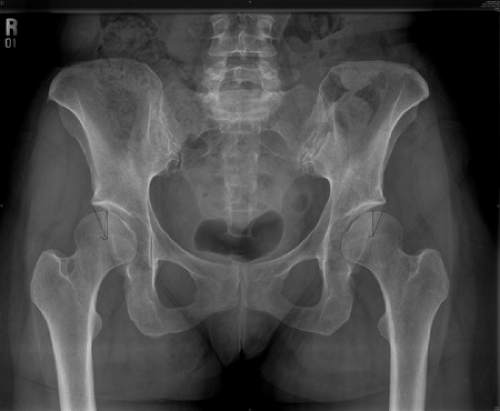 Figure 21A-2A Preoperative radiograph demonstrating hip dysplasia. |
Siebenrock and associates47 reported on Ganz’s experience on 71 hips with an average 11.3 year follow-up. At last follow-up, 82% had preservation of the hip joint, with 52 demonstrating good or excellent results and 6 with fair results using the Merle d’Aubigne clinical rating system. Prognostic indicators of poorer outcome included older age at time of operation, radiographic grade of arthritis, and presence of labral lesion. In review of their technique, the authors emphasize the importance of avoiding overcorrection of the pelvis as this may cause anterior femoroacetabular impingement.
Trousdale et al.48 described 42 patients followed for an average of 4 years after osteotomy. Thirty-two out of thirty-three patients with stage I or II arthritis had good to excellent results, while only eight out of nine patients with moderate to severe arthritic findings had Harris Hip Scores less than 70 or poor results. Six of these patients went on to THA. Clohisy and colleagues49 reported on 16 patients that underwent a Bernese periacetabular osteotomy for treatment of hip dysplasia. In the group, the average Harris Hip Score increased from 73.4 to 91.3 at an average of 4.2 years follow-up. Fourteen of the sixteen patients were satisfied with the results. There were two complications: loss of fixation requiring reoperation and overcorrection leading to ischial nonunion.
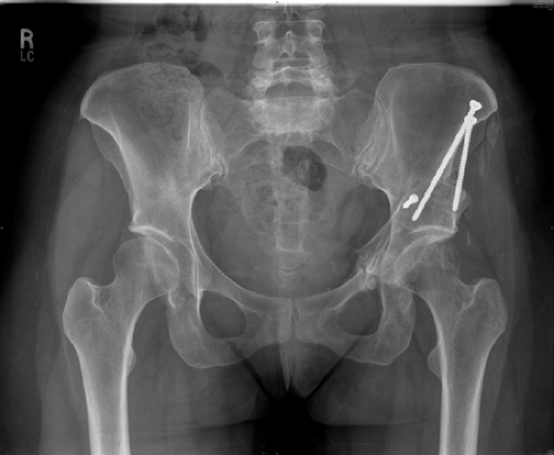 Figure 21A-2B Postoperative radiograph of Bernese periacetabular osteotomy. |
Salvage Pelvic Osteotomies
“Salvage” osteotomies such as the Chiari and the shelf osteotomy are performed in patients with severe dysplasia in whom joint congruency cannot be obtained with a reconstructive osteotomy. The Chiari osteotomy medializes the hip center producing a biomechanical advantage and provides better coverage of the femoral head. Windhager and associates50 reported on their series of 236 patients undergoing a Chiari osteotomy. Their results demonstrate that 215 patients did not need further surgery at a mean of 24.8 years. In this group, 51% had good results, 30% had fair results, and 18% had poor results. Other groups have documented somewhere between 60% and 75% clinical success at the latest follow-up.51,52,53 In summary, the authors concluded that 1) increased arthritis led to poorer results, 2) older patients did worse than younger ones, and 3) the results deteriorate with time.54
The Shelf procedure is described as corticocancellous augmentation to the anterolateral dome of the acetabulum in order to increase femoral head coverage. It is postulated that the bone augmentation undergoes metaplasia becoming a fibrocartilaginous structure. Migaud et al.55 reported on 56 shelf arthroplasties with an average 17 years follow-up. Survival at 20 years was 37%. When there was no preop evidence of arthrosis, the survivorship increased to 83% at 20 years. Furthermore, the patients that required conversion to THA had sufficient bone stock for conventional placement of the acetabular socket.
In summary, the most predictable indicator of outcome after osteotomy is the amount of arthritis present at the time of osteotomy. Therefore, it is important to identify the appropriate patients with dysplasia of the hip early. Patients who do not demonstrate significant radiographic evidence of arthrosis are ideal candidates. Results also correlate with achieving adequate correction of the deformity. Careful preoperative evaluation and surgical planning as well as skilled technical execution contribute to the likelihood of a successful osteotomy.
Femoral Osteotomy
Proximal femoral osteotomies can be performed in conjunction with an acetabular osteotomy or in isolation when the primary site of deformity is in the femur. Conditions that must be met to perform a femoral osteotomy includes 1) ability to perform a satisfactory correction of the deformity; 2) ability to maintain a satisfactory range of motion after the correction; and 3) joint congruency after correction. Furthermore, the surgeon must keep in mind that these patients may progress to THA and should try to avoid fragment translation, as this may require a second osteotomy at the time of arthroplasty.
The most common deformities of proximal femur/ femoral neck are valgus deformity, spherical femoral head, and slight acetabular dysplasia. Historically, the osteotomy is usually performed through the intertrochanteric region in order to minimize the nonunion rate. The most frequently used hardware is a blade plate which allows for rigid fixation and facilitates rotation and angulation. In addition to varus, the usual osteotomy is placed in slight extension in order to gain increased anterior femoral head coverage. Flexion osteotomies are generally performed to rotate out small anterior lesions secondary to osteonecrosis. Valgus intertrochanteric osteotomies are indicated with flattened femoral heads with a large medial femoral head osteophyte. A valgus osteotomy can decrease the joint reactive forces through medialization of the center of hip rotation, increased leg length, and improved abductor function.56 Patients should be warned about the leg length discrepancy that may occur.
The results of proximal femoral osteotomies have been varied. Varus osteotomies have produced better results than valgus osteotomies as these patients usually have mild dysplasia with minimal accompanied arthritis as described earlier. Iwase et al.57 reported on long-term results of both valgus and varus intertrochanteric osteotomies. In the varus group, survivorship from clinical failure was 89%, 87%, and 82% at 10, 15, and 20 years, respectively, while in the valgus group, the numbers were strikingly worse at 66%, 38%, and 19% at the same time points. In a meta-analysis of all current data regarding valgus and varus proximal femoral osteotomies, at 10 to 15 years, about 25% of varus osteotomies require hip arthroplasty, while nearly 50% of valgus osteotomies require arthroplasty.58,59 Today, proximal femoral osteotomies are performed less commonly, as these cases with acetabular dysplasia are usually addressed with an acetabular osteotomy.
Hip Joint Arthroplasty
History
THA remains the standard of care in the treatment of end stage arthrosis. Hip joint arthroplasty has been in a constant state of evolution in terms of implant design, biomaterials, and surgical technique for the past 40 years or more. Initial attempts to treat arthritic conditions interposed tissues (interpositional arthroplasty) between the worn articular surfaces.60 In the 1920s, Smith-Peterson introduced the concept of “mould arthroplasty.”61 This concept was spurred by an observation that he made when a piece of glass was embedded in the subcutaneous tissue of a patient. He noticed that “it was lined by a glistening synovial sac, containing a few drops of clear yellow fluid” and compared this to the normal synovial lining of the hip joint. The cup arthroplasty era began in 1923 when he implanted his first glass mold. His goal was to induce formation of a cartilaginous material, then to remove the glass mold. The initial attempts had problems with breakage of the glass mold which led to the experimentation and the development of Vitallium, a metal alloy.
The first total hip replacement is attributed to Phillip Wales.49 He implanted a stainless steel cup to the femoral neck that was fixed with a bolt and a similar sized stainless steel socket into the acetabulum fixed to the pelvis with screws. These large metal-to-metal articulations became dominant in use for many years. Sir John Charnley pioneered the modern era of THA with his work in bone
cement for fixation, introduction of polyethylene as a bearing material, use of aseptic techniques, and his diligence in recording patient follow-up.62,63,64,65
cement for fixation, introduction of polyethylene as a bearing material, use of aseptic techniques, and his diligence in recording patient follow-up.62,63,64,65
Hemiresurfacing and Total Hip Resurfacing
Hip resurfacing is a renewal of an old concept that began with the cup arthroplasty as described by Smith-Peterson and subsequent bone preserving arthroplasties. Hip resurfacing is theoretically an attractive option as a time buying procedure for young patients with disabling OA but no other factor that would limit physical activity.
Hemiresurfacing is a cemented hemispherical femoral head implant that is an option in patients without evidence of acetabular arthrosis. Clinical results from hemiresurfacing have not been reliable clinically as only approximately two thirds of patients experience pain relief.66,67 This is most likely related to implant articulating on intact acetabular cartilage.
Older generation total resurfacing was a failure due to volumetric polyethylene wear, but modern metal-on-metal articulations have demonstrated reduced wear and renewed interest in this implant. Mesko et al.68 described their experience with the older generation total articular replacement that included a femoral head resurfacing and a cemented all polyethylene acetabular component. From a total of 174 hips, 23 underwent revision surgery for component failure at an average of 5.1 years. Survivorship was 84.5% at 9 years. The reason for failure was high volumetric wear due to the large size of the femoral head. In addition, the large size of the femoral head led to a thin polyethylene as well as increased reaming of the acetabular bone stock in order to gain adequate fixation.
More recently, Amstutz et al.69 reported on 400 metal-on-metal total hip resurfacings performed on patients of an average age of 48 (Fig. 21A-3). The implant consisted of a porous-coated hemispherical acetabular component and a cemented femoral component that resurfaced the head. This system had the benefits of a low wear articulation, large diameter femoral head to reduce the chance for dislocation, and preservation of bone stock. Four-year survivorship has been 94%. No revisions have been performed due to the acetabular component; however, ten hips were revised to THA secondary to femoral component loosening or femoral neck fracture. The clinical results have been promising with an average Harris Hip Score of 93.5. Strict contraindications of this procedure include metaphyseal cysts (seen in osteonecrosis) and osteoporosis. The early results seem promising with reliable clinical results; however, longer follow-up is necessary to determine the durability of the prosthesis. In addition, patient selection has to be carefully defined to limit these early failures as total resurfacing is not as versatile as total hip replacement.
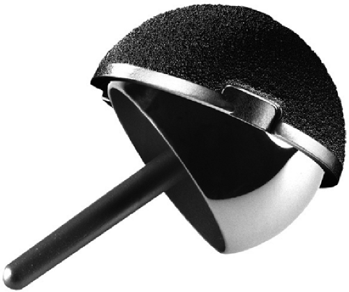 Figure 21A-3 Conserve Plus metal-on-metal total resurfacing prosthesis. |
Total Hip Arthroplasty
Epidemiology
The prevalence of THA has steadily increased between 1990 and 2002 according to the National Hospital Discharge Survey (NHDS).70 Over this time period, the rate of total hip replacement has increased nearly 50% per 100,000 patients. In contrast, total knee replacement has tripled. When comparing total hip revision to total knee revision surgery, the revision rate for hips (17.5%) was twice that of knees (8.2%).
In the health care environment today, health care costs and the ratio of cost and benefit are critically analyzed for every given procedure. Total hip replacement is one of the most beneficial surgical procedures that we currently perform.71,72 Barber and Healy73 reported that the actual cost of THA rose approximately 46.5% between 1981 and 1990. However, once adjusted for inflation, the actual increase was only 2%. When the components of the cost were examined, the cost of implants during this same time period had increased 212%, and the increase was 117% when adjusted for inflation. In 1981, the cost of the implant was 11% of the cost, while in 1990, the cost had become 24%. In order to maintain cost, surgical time has become more efficient, hospital stays have been shortened, and ancillary services have been employed more judiciously. Bozic et al.74 examined the actual costs of primary THA at their institution. The mean total costs were $24,170, with a mean hospital stay of 5.6 days. Worse preoperative medical health was a predictive factor of higher resource utilization.
Related posts:


Stay updated, free articles. Join our Telegram channel

Full access? Get Clinical Tree


